
Comparative Analysis of Germ Layer Composition in Teratomas Derived from Different Stem Cell Species
This study investigates the germ layer composition in teratomas derived from mouse, non-human primate, and human embryonic stem cells. Teratomas serve as key indicators of pluripotency, showing varying amounts of ectoderm, mesoderm, and endoderm based on the source of the stem cells. By conducting a semiquantitative histological analysis, our findings reveal significant species-specific differences in tissue organization within teratomas, suggesting underlying developmental programming variances. Further image analysis may enhance understanding of teratoma biology.

Comparative Analysis of Germ Layer Composition in Teratomas Derived from Different Stem Cell Species
E N D
Presentation Transcript
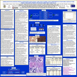
Figure 2 GENERIC CLASSIFIER GENERIC CLASSIFIER GLOBAL DECISION MR DECOMPOSITION GENERIC CLASSIFIER GENERIC CLASSIFIER IMAGE Figure 3 Figure 4 S1 S1 S1 S1 Figure 4: A) Original image without hue or saturation data. B) Image in A after 2-stage discrete wavelet transformation. WEIGHTING GENERIC CLASSIFIER INPUT IMAGE FEATURE EXTRACTOR CLASS LABEL CLASSIFIER Figure 5 Figure 1 Teratoma derived from nhpES cells with representative tissues derived from ectoderm: A) Neuroepithelium and B) skin. Tissues derived from mesoderm include C) bone and D) skeletal muscle. Tissues derived from endoderm are highlighted by E) respiratory epithelium and F) intestinal epithelium. SEMIQUANTITATIVE AND MULTIRESOLUTION-BASED HISTOLOGICAL ANALYSIS OF GERM LAYER COMPONENTS IN TERATOMAS DERIVED FROM HUMAN, NON-HUMAN PRIMATE AND MOUSE EMBRYONIC STEM CELLS. John A. Ozolek1, Carlos A. Castro2, Garrett Jenkinson3,4, Amina Chebira3, Jelena Kovacevic3,4, Christopher S. Navara2, Meena Sukhwani2, Kyle E. Orwig2, Ahmi Ben-Yehudah2, Gerald Schatten2 1Department of Pathology, Children's Hospital of Pittsburgh, University of Pittsburgh, Pittsburgh, PA, USA 2Department of Obstetrics and Gynecology, Magee Womens Research Institute and Foundation, University of Pittsburgh, Pittsburgh, PA, USA 3Department of Biomedical Engineering, Carnegie Mellon University, Pittsburgh, PA, USA4Department of Electrical and Computer Engineering, Carnegie Mellon University, Pittsburgh, PA, USA ABSTRACT Background: The capability of cells derived as embryonic stem cells (ES) to produce tissue components comprising the three developmental germ layers (teratomas) is the single most important test of pluripotency. Within the pathology literature, teratomas have been classified according to the complexity of tissue organization such that in diagnostic terms, lesions with two of the three germ layers are considered teratomas and lesions with high order arrangement of tissues resembling an embryo or fetus are considered by some to be teratomas. Little is known about the volume of distinct tissue types produced, how tissue types are organized, variables that may influence tissue differentiation, and species differences within teratomas. We hypothesize teratomas derived from ES cells of different mammalian species will exhibit species specific three dimensional tissue distribution and volumes within teratomas. Methods: Testes of SCID mice were injected with putative ES cells derived from mouse (MES) and non-human primate (nhpES). A human ES line (H7) was also used. Animals were sacrificed when visible lesions were identified. The entire teratoma was extracted, fixed in formalin, serially sectioned and processed by routine histological techniques. For each lesion, the amount of each representative germ layer (ectoderm (EC; neuroglial, skin), mesoderm (ME; mesenchyme, bone, cartilage), endoderm (EN; gastrointestinal, bronchial, pancreas)) was semiquantified according to the following scale: 1-0-20%, 2-21-40%, 3-41-60%, 4-61-80%, 5-81-100%. %germ layer is given as mode of percentage. Incubation times are given in days with standard deviation in parenthesis. Statistical analyses were done with ANOVA and t-test for continuous variables and Wilcoxon and Mann-Whitney tests for non-parametric variables. Results are expressed as (human vs nhp vs mouse) where values are given. Results: Days of ES cell incubation after mice injection were not statistically significant between groups (71 vs 78 vs 68). However, human ES derived teratomas were larger than nhpES teratomas, but not larger than MES teratomas (2.6 cm vs 1.8 cm vs 1.9 cm). Teratomas derived from MES and nhpES showed significantly higher amounts of EC (5 vs 1) than human ES teratomas while human derived teratomas demonstrated higher amounts of ME than nhp or mouse (4 vs 1). EN amount did not differ between groups. Within species nhpES and MES derived teratomas had greater amounts of EC than ME or EN while human derived teratomas had greater amounts of ME than EC or EN. Conclusions: We conclude that species differences exist by amounts of the various germ layers produced in teratomas and may not be related to incubation time or tumor size. We speculate that this may reflect basic developmental programming differences between the species. Further sophisticated bioimaging analysis and three-dimensional reconstruction of teratomas will further elucidate these differences.: • METHODS • Semiquantitative Analysis • Testes of NOD-SCID mice (Jackson Laboratories, Bar Harbor, Maine) were injected with putative ES cells derived from mouse (MES), non-human primate (nhpES [60,000-120,000cells/testes]), and a human (hES;H7 [3,000-4,500 cells/testes]) source. A total of 8, 3, and 2 teratomas were derived from non-human primate, murine, and human ES cells respectively. • Mice were sacrificed when visible lesions were identified. • The entire teratoma was carefully dissected and removed in its entirety and fixed in 10% phosphate buffered formalin (3.6% formaldehyde). • After fixation, lesions were measured, serially sectioned and processed by routine histological methods. • For each lesion, the amount of each representative germ layer (i.e. ectoderm, mesoderm, and endoderm) was estimated on each slide of the serially sectioned teratoma using the following scale: 1-[0-20%], 2-[21-40%], 3-[41-60%], 4-[61-80%], and 5-[81-100%]. Tissue components of each germ layer were identified according to the following table (Table 1). • Size (greatest dimension) of lesions is given in centimeters. Incubation times are given in days. The percentage of germ layer present is given as median of percentage. Statistical analyses were done with ANOVA and t-test for continuous variables and Wilcoxon and Mann-Whitney tests for non-parametric variables. Figure 6 Figure 2: Multiresolution classifier where an image is “decomposed” (see Figure 3) and the resultant subbands subjected to the generic classifier (Figure 1) until assigned class label (global decision) is achieved. SWT transformation provides highest accuracy of identifying specific tissue types compared to using DWT or the generic classifier alone. • CONCLUSIONS • In general, ectoderm and mesoderm predominate within teratomas derived from ES cells regardless of the species. • Teratomas derived from hES cells tend to be larger than those derived from nhpES or MES cells. • Non-human primate and mouse teratomas show a greater percentage of ectoderm derived tissue than human teratomas. Human teratomas show a greater percentage of mesoderm than non-human primate or mouse. For all species endoderm derived tissues are present in the least amount. • Using the multiresolution classifier with texture features only computed on the multiresolution-decomposed digital images of tissue types within teratomas, we obtain accuracy of 83%. • SPECULATIONS • We speculate that developmental programs and/or timing differ between species such that mammals with shorter gestational ages show greater prevalence of ectoderm derived tissues (particularly neural tissue which is first to develop) even in a seemingly disorganized conglomerate of tissues comprising the teratoma. • The ability to recognize and quantify tissue types using digital imaging analysis tools including MR transforms and then reconstruct these lesions in three dimensions will allow us to understand the spatial relationships of tissue types and correlate with high-resolution imaging studies. • Higher accuracy of tissue typing using these and other MR transforms can be achieved using color, shape, and location. • RESULTS • Semiquantitative Analysis • No differences were seen for incubation days between teratomas derived from nhpES, MES, or HES. The human teratomas sampled were significantly larger than either nhp or mouse derived lesions (Table 2). • Both nhp and mouse derived teratomas demonstrated higher median percentage of ectoderm derived tissue present in their teratomas compared to hES derived teratomas. However, hES cell derived teratomas demonstrated higher percentages of mesoderm derived tissues than nhpES or MES derived teratomas (Figure 5). No differences were seen for percentage of endoderm derived tissues between nhp and mouse ES teratomas. Significant differences at a p-value of 0.02 were seen between endoderm derived tissues from nhpES and MES compared to hES teratomas (Table 2). • Classification with and without Multiresolution • For tissue types selected for analysis (mesenchyme, skin, myenteric plexus, bone, necrosis, and striated muscle), the multiresolution classifier improved accuracy of detecting a particular tissue type over the use of a generic classifier. Stationary wavelet transform produced a mean of 83% accuracy compared to 75% and 68%, for discrete wavelet transform and no multiresolution respectively (Figure 6; Table 3). • INTRODUCTION • The ability to form lesions that recapitulate the three germ layers ectoderm, mesoderm, endoderm) during development is one of the assays (considered a gold standard) for determining if potential embryonic stem cell (ES cells) candidates are pluripotent. • Within the pathology literature, human teratomas are classified according to the presence of immature and/or malignant tissue elements as these have prognostic significance in the pediatric and adult populations. • While at first glance, most teratomas derived from ES cells appear as disorganized tissue masses with recognizable germ layer elements, little is known about the contribution of each germ layer to the lesion, the spatial organization of germ layer elements to one another, three-dimensional hierarchy of germ layer contribution and whether the “final” constitution of the teratoma is time and/or species dependent reflecting attempts to follow a developmental program. • The ability to accurately detect and quantify specific tissue types will begin to allow the ability to detect species specific differences in developmental programming and enable accurate three-dimensional reconstruction of teratomas and comparison to high-resolution magnetic resonance imaging. Discrete wavelet transform (DWT) with two levels of decomposition and reconstrcution. g and h are orthogonal lowpass and highpass filters • METHODS • Multiresolution Classification • Multiresolution techniques have been developed over 20 years ago. • Multiresolution classification new---First attempt to apply it to this type of data. • If feasible, will allow accurate classification and quantification of tissue types throughout the entire teratoma. • Results can be correlated with three-dimensional high-resolution magnetic resonance image renderings. • Generic classifier: Typically feature extraction (numerical) followed by classification (Figure 1). • Large multi-class images are separated into small single class images. • Texture features are used in neural net classifier using 10-fold cross validation. • Classification of tissue type achieved through multiresolution classification (Figure 2). It uses multiresolution decomposition---discrete wavelet transformation (DWT) or stationary wavelet transformation (SWT) (Figures 3, 4), followed by texture feature classification as well as weighting to combine local decisions into a global one. TABLE 1 Tissue components of germ layers TABLE 2 % of tissue types in species-specific teratomas • AIMS • Compare using a semiquantitative approach the contribution of each germ layer to teratoma formation within species and between species. • Determine whether germ layer contribution is based on the size of the teratoma or incubation time. • Determine the accuracy of multiresolution based imaging analysis techniques in identifying specific tissue types derived from each germ layer. • CORRESPONDENCE • John A. Ozolek, M.D. • Assistant Professor of Pathology • Children's Hospital of Pittsburgh • 3705 Fifth Avenue • Pittsburgh, PA 15213 • 412-692-5641/412-251-2248 (office/cell) • 412-692-5650 (Department) • 412-692-6550 (fax) • ozolekja@upmc.edu • Carlos A. Castro, D.M.D., M.D. • Research Associate • Department of Obstetrics, Gynecology and Reproductive Medicine • Magee Womens Research Institute • 204 Craft Avenue • Pittsburgh, PA 15213 • 412-641-6086/412-310-3091 (office/cell) • 412-641-2410 (fax) • pdccac@pdc.magee.edu SD in ( ), *-p<0.01, See text in Results section for statistical analysis of median percentage of germ layers TABLE 3 Accuracy of tissue classification using a multiresolution classifier Neural network based generic classifier where image features (texture, shape, color) are subjected to neural network until output matches desired class label


