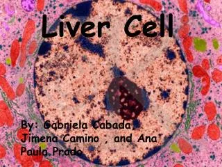
LIVER CELL FAILURE
LIVER CELL FAILURE. DONE BY : Fatima S. Bajari Ohood Al Bajri Malath Al Shereef. OBJECTIVES. Liver Anatomy Physiology Liver Cirrhosis. Liver Anatomy . liver is the largest gland of the body.


LIVER CELL FAILURE
E N D
Presentation Transcript
LIVER CELL FAILURE DONE BY : Fatima S. Bajari Ohood Al Bajri Malath Al Shereef
OBJECTIVES • Liver Anatomy • Physiology • Liver Cirrhosis
Liver Anatomy • liver is the largest gland of the body. • the liver is divided into right and left lobes by the external marking of the falciform ligament.
Liver Segments II VIII IV III VII • composed of 4 lobes (left, right, caudate, quadrate) and divided into 8 segments IV V VI The Right Lobe The Left Lobe • Each segment receives its own portal pedicle (triad of portal vein, hepatic artery, and bile duct).
Blood Supply Portal v. 70% Hepatic a. 30%
Liver Physiology the liver performs several important functions including: • Protein metabolism (Synthesis and storage) • Carbohydrate metabolism • Lipid metabolism • Formation of bile (Bile secretion and bile acid metabolism) • Hormone and drug inactivation • Immunological function
Protein metabolism (Synthesis and storage) • The liver is the principal site of synthesis of all circulating proteins, which are produced in the reticuloendothelialsystem. • Plasma contains 60–80 g/L of protein, mainly in the form of albumin, globulin and fibrinogen . • Albumin main functions are first to maintain the intravascular oncotic (colloid osmotic) pressure, and second to transport water-insoluble substances such as bilirubin,hormones, fatty acids and drugs. • Transport or carrier proteins such as transferrin and caeruloplasmin. • The liver also synthesizes all factors involved in coagulation ( fibrinogen, prothrombin, factors V, VII, IX, X and XIII, proteins C and S and antithrombinas well as components of the complement system) • The liver stores large amounts of vitamins, particularly A, D and B12, vitamin K and folate), and also minerals – iron in ferritin and haemosiderinand copper. • As The liver receives amino acids degraded by transamination and oxidative deamination ammonia, convertedto urea excreted by kidneys. Liver Physiology Cont…
Carbohydrate metabolism • Glucose homeostasis and the maintenance of the blood sugaris a major function of the liver. • In the immediate fasting state, blood glucose is maintained either by glucose released from the breakdown of glycogen (glycogenolysis) or by newly synthesized glucose (gluconeogenesis). • Sources for gluconeogenesis are lactate, pyruvate, amino acids from muscles (mainly alanine and glutamine) and glycerol from lipolysis of fat stores. • In prolonged starvation, ketone bodies and fatty acids are used as alternative sources of fuel as the body tissues adapt to a lower glucose requirement Liver Physiology Cont…
Lipid metabolism • Fats are insoluble in water and are transported in the plasma as protein–lipid complexes (lipoproteins). • The liver has a major role in the metabolism of lipoproteins. It synthesizes (VLDLs) and (HDLs). • Hepatic lipase removes triglyceride from (IDLs) to produce (LDLs) which are degraded by the liver after uptake by specific cell receptors. • Oxidation of FFA occurs in the liver, depending on the availability of dietary fat .Cholesterol may be of dietary origin but most is synthesized from acetyl-CoA mainly in the liver, adrenal cortex and skin. Liver Physiology Cont…
Formation of bile (Bile secretion and bile acid metabolism) - RBCs >> heme Oxidized into >> bileverdine >> reduction to (indirect bilirubin). 1- Uptake of indirect bilirubin through Y and Z receptors in the liver. 2- Conjugation and Secretion: - Conjugationoccurs in the liver by glucouronyltransfereaseand the conjugated bilirubin is secreted with the bileinto the small intestine in the form of stercobilinogen which passing into three directions: • Part of it enters the enterohepaticcirculation. • Another part escapes to the systemic circulation reaching the kidneyand secreted in urine (urobilinogen). • The last part passes to the large intestineto be transformed into stercobilin. Liver Physiology Cont…
Hormone and drug inactivation • The liver catabolizes hormones such as insulin, glucagon, estrogens, growth hormone, glucocorticoids and parathyroid hormone. • It is also the prime target organ for many hormones (e.g. insulin). It is the major site for the metabolism of drugs and alcohol . • Fat-soluble drugs are converted to water-soluble substances that facilitate their excretion in the bile or urine. • Cholecalciferol is converted to 25-hydroxycholecalciferol. Liver Physiology Cont…
Immunological function • The reticuloendothelial system of the liver contains many immunologically active cells. • The liver acts as a sieve for the bacterial and other antigens carried to it through the portal vein from G.I.T., they are phagocytosed by Kupffer cells, these cells secrete interleukins and tumour necrosis factor (TNF). • The reticuloendothelialsystem plays a role in tissue repair, T and B lymphocyte interaction, and cytotoxic activity in disease processes. Liver Physiology Cont…
Liver Cirrhosis Definition: • It is a chronic liver disease results from necrosis of hepatocytes followed by fibrous tissue deposition and formation of regenerating nodules with loss of hepatic architecture. • This derangement eventually produces portal hypertension and liver cell failure.
Aetiology • Alcohol is now the most common cause in the West, but viral infection is the most common cause world-wide. • Young patients with cirrhosis must be investigated carefully as the cause may be treatable (e.g.Wilson’s disease).
Pathogenesis • Long standing injury to the liver lead to inflammation, necrosis and eventually fibrosis(initiated by activation of stellate cells). • These liver injuries e.g. (virus, alcohol, .... ) stimulate kupffer cells release of cytokines stimulate into (stellate) cells excessive release and deposition of collagen fibresloss of hepatic architecture (cirrhosis).
Pathology The characteristic features of cirrhosis are regenerating nodules separated by fibrous septa and loss of the normal lobular architecture within the nodules . Two types of cirrhosis have been described which give clues to the underlying cause: ■Micronodular cirrhosis: Regenerating nodules are usually less than 3 mm in size and the liver is involved uniformly. This type is often caused by ongoing alcohol damage or biliary tract disease. ■ Macronodular cirrhosis.: The nodules are of variable size and normal acini may be seen within the larger nodules. This type is often seen following chronic viral hepatitis. A mixed picture with small and large nodules is sometimes seen.
Investigations • These are performed to assess the severity and type of liver disease. 1- Severity ■ Liver function: Serum albumin and prothrombin time are the best indicators of liver function ■ Liver biochemistry: This can be normal, depending on the severity of cirrhosis. In most cases there is at least a slight elevation in the serum ALP and serum aminotransferases. ■ Serum electrolytes: A low sodium indicates severe liver diseasedue to a defect in free water clearance or to excess diuretic therapy. ■ Serum creatinine: An elevated concentration > 130 μmol/L is a marker of worse prognosis. In addition, serum α-fetoprotein if > 200 ng/mL is strongly suggestive of the presence of a hepatocellular carcinoma.
Cont. Investigations 2- Type This can be determined by: ■ viral markers ■ serum autoantibodies ■ serum immunoglobulins ■ iron indices and ferritin (Total iron-binding capacity (TIBC) and ferritin should be measured to exclude hereditary Haemochromatosis) ■ copper, caeruloplasmin (Serum copper and serum α1-antitrypsin should always be measured in young cirrhotics. ) ■ α1-antitrypsin ■ Ultrasound examination: This can demonstrate changes in size and shape of the liver. Fatty change and fibrosis produce a diffuse increased echogenicity. The patency of the portal and hepatic veins can be evaluated. It is useful in detecting hepatocellular carcinoma ■ CT scan ■ Endoscopy is performed for the detection and treatment of varices, and portal hypertensive ■ Biopsy: This is usually necessary to confirm the severity and type of liver disease.
Management • Patients should have 6-monthly ultrasound to detect the early development of a hepatocellular carcinoma. • Treatment of the underlying cause may arrest or occasionally reverse the cirrhotic changes . • Patientswith compensated cirrhosis should lead a normal life. • Theonly dietary restriction is to reduce salt intake. • Aspirin and NSAIDs should be avoided. • Alcohol should be avoided ??
LIVER TRANSPLANTATION What are the Indications? - Acute liver disease: Patients with fulminant hepatic failure of any cause, including acute viral hepatitis may be considered. - Chronic liver disease: The indications for transplantation are usually for complications of cirrhosis, no longer responsive to therapy. - All patients with end-stage (Child’s grade C) cirrhosis. . In addition specific extrahepatic complications of cirrhosis, even with preserved liver function, such as hepatopulmonary syndrome (shunting in the lung leading to hypoxia) and porto-pulmonary hypertension,can be reversed by liver transplantation. - Primary biliary cirrhosis: Patients with this disease should be transplanted when their serum bilirubin is persistently > 100 μmol/L or symptoms such as itching are intolerable. - Chronic hepatitis B if HBV DNA negative or levels falling under therapy. Following transplantation, recurrence of hepatitis is prevented by hepatitis B immunoglobulin
LIVER TRANSPLANTATION Cont.. - Chronic hepatitis C is the most common indication. • Autoimmune hepatitis. In patients who have failed to respond to medical treatment or have major side-effects of corticosteroid therapy. • Alcoholic liver disease !! • Primary metabolic disorders. Examples are Wilson’s disease, hereditary haemochromatosis and α1- antitrypsin deficiency. • Other conditions, such as sclerosingcholangitis
LIVER TRANSPLANTATION Cont.. Contraindications: Absolute contraindications: • active sepsis outside the hepatobiliary tree • malignancy outside the liver, • liver metastases • If the patient is not psychologically committed. Relative contraindications: • mainly anatomical considerations that would make surgery more difficult, such as extensive splanchnic venous thrombosis. • With exceptions, patientsaged 65 years or over are not usually transplanted. • In hepatocellular carcinoma the recurrence rate is high unless there are fewer than three small (< 3 cm) lesions, or a solitary nodule of < 5 cm.
COMPLICATIONS AND EFFECTS OF CIRRHOSIS Portal hypertension Varicealhaemorrhage Ascites Portosystemic encephalopathy (PSE) Renal failure (hepatorenal syndrome) Hepatopulmonary syndrome Porto-pulmonary hypertension Primary hepatocellular carcinoma
Portal hypertension • The portal vein is formed by the union of the superior mesenteric and splenic veins. • The pressure within it is normally 5–8 mmHg with only a small gradient across the liver to the hepatic vein in which blood is returned to the heart via the inferior vena cava. • Portal hypertension can be classified according to the site of obstruction: • ■ prehepatic – due to blockage of the portal vein before the liver • ■ intrahepatic – due to distortion of the liver architecture, which can be presinusoidal (e.g. in schistosomiasis) or postsinusoidal (e.g. in cirrhosis) • ■ posthepatic – due to venous blockage outside the liver(rare). Cont. complications and effects of cirrhosis: 1-Portal hypertension
Portal hypertension • As portal pressure rises above 10–12 mmHg the compliant venous system dilates and collaterals occur within the systemic venous system. • The main sites of the collaterals are at the gastro-oesophageal junction, therectum, theleft renal vein, thediaphragm, theretroperitoneumand the anterior • abdominal wall via the umbilical vein. • The collaterals at the gastro-oesophageal junction (varices) • Rectal varices are found frequently (30%) • gut becomes congested giving rise to portal hypertensive gastropathy and colopathy, in which there is punctateerythema sometimes erosions, which can bleed. Cont. complications and effects of cirrhosis: 1-Portal hypertension
Pathophysiology • Portal vascular resistance is increased in chronic liver disease. • During liver injury: stellate cells are activated and transform into myofibroblasts. • expression of the specific smooth muscle protein α-actin under the influence of endothelin, nitricoxide or prostaglandins. • the contraction of these activated cells contributes to abnormal blood flow patterns and increased resistance to blood flow. • the balance of fibrogenic and fibrolytic factors is shifted towards fibrogenesis. • leads to portal hypertension and opening of portosystemicanastomoses in both precirrhotic and cirrhotic livers. Neoangiogenesis also occurs. • Patients with cirrhosis have a hyperdynamic circulation due to the release of nitric oxide and glucagon, which leads to peripheral and splanchnic vasodilatation. • This effect is followed by plasma volume expansion due to sodium retention and this has a significant effect in maintaining portal hypertension. Cont. complications and effects of cirrhosis: 1-Portal hypertension
Causes Prehepatic causes Intrahepatic causes Cont. complications and effects of cirrhosis: 1-Portal hypertension Posthepatic causes
Prehepatic causes • Extrahepatic blockage is due to portal vein thrombosis. • The cause is often unidentified, but some cases are due to portal vein occlusion secondary to congenital portal venous abnormalities or neonatal sepsis of the umbilical vein. • Many are due to inherited defects causing prothrombotic conditions, e.g. factor V Leiden. • Patients usually present with bleeding, often at a young age. • They have normal liver function and, prognosis excellent. • The portal vein blockage can be identified by ultrasound with Doppler imaging; CT and MR angiography. • Treatment is repeated endoscopic therapy or nonselective beta-blockade. • Splenectomy is only performed if there is isolated splenic vein thrombosis. • Anticoagulation prevents further thrombosis and does not increase the risk • of bleeding; used when there is a high risk of recurrent thrombosis. Cont. complications and effects of cirrhosis: 1-Portal hypertension
Intrahepatic causes cirrhosis is the most common intrahepatic cause of portal hypertension other causes: ■ Non-cirrhotic portal hypertension. portal hypertension and variceal bleeding without cirrhosis. Histologically, the liver shows mild portal tract fibrosis. The aetiology is unknown, but arsenic, vinyl chloride, antiretroviral therapy and other toxic agents have been implicated. A similar disease is found frequently in India. liver lesion does not progress and the prognosis is therefore good. ■Schistosomiasiswith extensive fibrosis is the commonest cause, endemic areas such as Egypt and Brazil. often there may be concomitant liver disease such as HCV infection . ■ Other causes include congenital hepatic fibrosis, nodular regenerative hyperplasia and partial nodular transformation(rare). The common features of hyperplastic liver cell growth in the form of nodules but in contrast to cirrhosis, fibrosis is typically absent. A wedge liver biopsy is usually required to establish the diagnosis. hormones are none implicated in aetiology or progression. Cont. complications and effects of cirrhosis: 1-Portal hypertension
Posthepatic causes • Prolonged severe heart failure with tricuspid incompetence • constrictive pericarditis Cont. complications and effects of cirrhosis: 1-Portal hypertension both lead to portal hypertension.
Clinical features Patients with portal hypertension are often asymptomatic and the only clinical evidence of portal hypertension is splenomegaly. Clinical features of chronic liver disease are usually present. Presenting features may include: ■ haematemesis or melaena from rupture of gastro-oesophagealvarices or portal hypertensive gastropathy ■ ascites ■ encephalopathy ■ breathlessness due to porto-pulmonary hypertension or hepatopulmonary syndrome (rare). Cont. complications and effects of cirrhosis: 1-Portal hypertension
Variceal haemorrhage • Approximately 90% of patients with cirrhosis will develop gastro-oesophagealvarices, over 10 years, only onethird of these will bleed from them. • Bleeding is likely to occur with large varices, red signs on varices (diagnosed at endoscopy) and in severe liver disease. Cont. complications and effects of cirrhosis: 2-Variceal haemorrhage
Management • Management can be divided into the active bleeding episode,theprevention of rebleeding, and prophylactic measures to prevent the first haemorrhage. • the prognosis depends on the severity of the underlying liver disease, with an overall mortality from varicealhaemorrhage of 25%, reaching50% in Child’s grade C. Cont. complications and effects of cirrhosis: 2-Variceal haemorrhage
Initial management of acute variceal bleeding Cont. complications and effects of cirrhosis: 2-Variceal haemorrhage
Resuscitation ■ Assess the general condition of the patient – pulse and blood pressure. ■ Insert an intravenous line and obtain blood for grouping and crossmatching, haemoglobin, PT/INR, urea, electrolytes, creatinine, liver biochemistry and blood cultures. ■ Restore blood volume with plasma expanders or blood transfusion. Prompt correction of hypovolaemia is necessary in patients with cirrhosis as their baroreceptor reflexes are diminished. ■ Ascitic tap. ■ Monitor for alcohol withdrawal. Give thiamine i.v. ■ Start prophylactic antibiotics – third generation cephalosporins, e.g. cefotaxime. These treat and prevent infection and early rebleeding and reduce mortality. Cont. complications and effects of cirrhosis: 2-Variceal haemorrhage
Urgent endoscopy • toconfirm the diagnosis of varices. • excludes bleeding from other sites (e.g. gastric ulceration) or portal hypertensive (or congestive) gastropathy. • injected with a sclerosing agent that may arrest bleeding by producing vessel thrombosis Cont. complications and effects of cirrhosis: 2-Variceal haemorrhage Balloon tamponade is used mainly to control bleeding if endoscopic therapy or vasoconstrictor therapy has failed Transjugular intrahepatic portocaval shunt (TIPS) is used when bleeding cannot be stopped after two sessions of endoscopic therapy within 5 days. Emergency surgery This is used when other measures fail or if TIPS is not available and, particularly, if the rebleeding is from gastric fundal varices.
Long-term measures Oral propranolol in a dose sufficient to reduce resting pulse rate by 25% has been shown to decrease portal pressure. Portal inflow is reduced by two mechanisms: decrease in cardiac output (β1), blockade of β2 vasodilator receptors on the splanchnic arteries, leaving an unopposed vasoconstrictor effect. Cont. complications and effects of cirrhosis: 2-Variceal haemorrhage
Ascites The pathogenesis of ascites in liver disease is secondary to renal sodium and water retention. Several factors are involved: ■ Sodium and water retention results from peripheral arterial vasodilatation and consequent reduction in the effective blood volume. ■ Portal hypertension exerts a local hydrostatic pressure and leads to increased hepatic and splanchnic production of lymph and transudation of fluid into the peritoneal cavity. ■ Low serum albumin (a consequence of poor synthetic liver function) may further contribute by a reduction in plasma oncotic pressure. Cont. complications and effects of cirrhosis:3- Ascites
Clinical features The abdominal swelling associated with ascitesdevelops over many weeks or as rapidly as a few days. Precipitating factors include a high sodium diet or the development of a hepatocellular carcinoma or splanchnic vein thrombosis. Mild generalized abdominal pain and discomfort are common. Respiratory distress accompanies tense ascites, and also causes difficulty in eating. confirmed by the demonstration of shifting dullness also have peripheral oedema. A pleural effusion (usually on the right side) may infrequently be found and arises from the passage of ascitic fluid through congenital diaphragmatic defects. Cont. complications and effects of cirrhosis:3- Ascites
Investigations A diagnostic aspiration of 10–20 mL of fluid should beobtained and the following performed: ■Cell count. A neutrophil count above 250 cells/mm3 is indicative of an underlying (usually spontaneous) bacterial peritonitis. ■Gram stain and culture – for bacteria and acid-fast bacilli. ■Protein. A high serum–ascites albumin gradient of > 11 g/L suggests portal hypertension, and a low gradient < 11 g/L is associated with abnormalities of the peritoneum, e.g. inflammation, infections, neoplasia ■Cytology – for malignant cells. ■ Amylase – to exclude pancreatic ascites Cont. complications and effects of cirrhosis:3- Ascites
Management The aim is to reduce sodium intake and increase renal excretion of sodium, ■ Check serum electrolytes and creatinineat the start and every other day; weigh patient and measure urinary output daily. ■ Bed rest alone will lead to a diuresis in a small proportion of people by improving renal perfusion, but in practice is not helpful. ■ By dietary sodium restriction it is possible to reduce sodium intake to 40 mmol in 24 hours and still maintain an adequate protein and calorie intake with a palatable diet. ■ Drugs: many contain significant amounts of sodium (up to 50 mmol daily). include antacids, antibiotics (particularly the penicillins and cephalosporins) and effervescent tablets. Sodium-retaining drugs (nonsteroidals, corticosteroids) should be avoided. Cont. complications and effects of cirrhosis:3- Ascites
Cont. Management ■ Fluid restriction is probably not necessary unless the serum sodium is under 128 mmol/L. ■ The diuretic of first choice is the aldosterone antagonist spironolactone ■ ParacentesisThis is used to relieve symptomatic tense ascites. ■ Shunts a transjugularintrahepaticportosystemic shunt (TIPS) is used for resistant ascites providing there is no spontaneous portosystemic encephalopathy and minimal disturbance of renal function. Cont. complications and effects of cirrhosis:3- Ascites
Spontaneous bacterial peritonitis (SBP) • A serious complication of ascites with cirrhosis. • occurs in approximately 8%. The infecting. • Organisms access to the peritoneum by haematogenous spread • most are Escherichia coli, Klebsiella or enterococci. • The condition should be suspected in any patient with ascites who clinically deteriorates. • A raised neutrophil count in ascites is alone sufficient evidence to start treatment • immediately. • A third-generation cephalosporin (cefotaxime or ceftazidime) is used and is modified on the basis of culture results. • Mortality is 10–15%. • Recurrence is common (70% within a year) and an oral quinolone • SBP is an indication to refer to a liver transplant centre. Cont. complications and effects of cirrhosis:3- Ascites