Acid-Base Disturbances in Metabolic Processes
370 likes | 403 Vues
Learn about the production of acid and base in metabolic processes, buffer systems, pulmonary and renal regulation, causes of respiratory acidosis, metabolic effects, and treatment principles for acid-base imbalances.

Acid-Base Disturbances in Metabolic Processes
E N D
Presentation Transcript
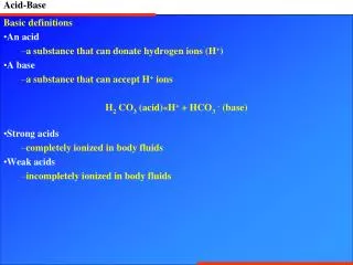
Metabolic processes continually produce acid and, to a lesser degree, base. • Small amounts (40-80mmol/24h) are formed from the oxidation of amino acids and the anaerobic metabolism of glucose to lactic and pyruvic acid. • Far more acid is produced as a result of carbon dioxide (CO2) release from oxidative (aerobic) metabolism - 15,000mmol/24h (15x103 mmol/24h). • CO2 rapidly reacts with water to form carbonic acid (H2CO3), which further dissociates into hydrogen and bicarbonate ions (HCO3-).
Enzymes function optimally over a very narrow range of hydrogen ion concentrations. For most enzymes this optimum pH is close to the physiological range for plasma (pH= 7.35 to 7.45).
Figure shows a typical graph obtained when enzyme activity is plotted against pH. The curve is a narrow bell shape centred around physiological pH.
Buffer • A buffer is a solution containing substances which have the ability to minimise changes in pH when an acid or base is added to it . • A buffer typically consists of a solution which contains a weak acid mixed with the salt of that acid & a strong base.
Buffer systems • a) Bicarbonate • b) Proteins • c) Haemoglobin • d) The phosphate • buffer systems act within seconds
Bicarbonate buffer • Most acids are neutralized by basic component. • Bases are neutralized by acid component.
Pulmonary regulation: • ↑ H+ ------ ↑ H2CO3 • H2CO3 → H2O + CO2↑ TV RR • ↓ H+ ------ ↓H2CO3↓ TV RR • Respiratory mechanisms responds within minutes, maximal in 12-24 hrs.
Renal regulation • Reabsorption of Bicarbonate b) Excretion of Hydrogen Ions c) Ammonia generation • Renal mechanisms responds slowly (effectively in 3-5 days)
Respiratory acidosis • Respiratory Acidosis is an acid base disturbance characterized by an elevation in the partial pressure of CO2 leading to an increase in the hydrogen ion concentration • [H+] = 24 × PCO2 / [HCO3-]
Respiratory acidosis • Causes of respiratory acidosis: • CNS depression 1. Opioids 2. Oxygen in patient with chronic hypercapnia 3. Central sleep apnea 4. CNS lesion 5. Extreme obesity (Pickwickian syndrome)
Respiratory acidosis • Chest wall or Thoracic Cage Abnormality 1. Kyphoscoliosis 2. Flail Chest 3. Myxedema 4. Rib Fracture 5. Scleroderma • Neuromuscular disorders 1. Myasthenia gravis 2. Guillain-Barre 4. Poliomyelitis 5. Muscular dystrophy 6. Multiple Sclerosis
Respiratory acidosis • Disorders affecting gas exchange 1. COPD 2. Severe asthma or pneumonia 3. Pneumothorax or Hemothorax 4. Acute pulmonary edemaAirway obstruction 1. Aspiration of foreign body 2. Obstructive sleep apnea 3. Laryngospasm
Metabolic Effects • Stimulation of ventilation via both central and peripheral chemoreceptors • Cerebral vasodilation increasing cerebral blood flow and intracranial pressure • Stimulation of the sympathetic nervous system resulting in tachycardia, peripheral vasoconstriction and sweating • Peripheral vasodilation by direct effect on vessels • Central depression at very high levels of pCO2
TREATMENT • Treat underlying disorder • Supply oxygen • Corticosteroids and bronchodilators to reduce airway inflammation and resistance. • Mechanical ventilator if ventilation fails.
Metabolic Acidosis A metabolic acidosis is an abnormal primary process or condition leading to an increase in fixed acids in the blood.
Causes of Metabolic Acidosis • Increased Endogenous production: Ketoacidosis (Alcohol, Starvation, DKA) Lactic AcidosisUremia Intoxications: Methanol, Ethylene Glycol, Salicylates Loss of Bicarbonate: Diarrhea Pancreatic, biliary, intestinal fistula Exogenous Administration: HCL Decreased Renal Acid Excretion: Renal Failure
Respiratory Effects • Hyperventilation ( Kussmaul respirations) - this is the compensatory response • Shift of oxyhaemoglobin dissociation curve (ODC) to the right • Decreased 2,3 DPG levels in red cells (shifting the ODC back to the left)
Cardiovascular effects: • Depression of myocardial contractility • Sympathetic overactivity (incl tachycardia, vasoconstriction,decreased arrhythmia threshold) • Resistance to the effects of catecholamines • Peripheral arteriolar vasodilatation • Venoconstriction of peripheral veins • Vasoconstriction of pulmonary arteries • Effects of hyperkalaemia on heart
Treatment Principles 1. Emergency management of immediately life-threatening conditions i.e. intubation and ventilation for airway or ventilatory control; CPR. 2. Treat the underlying disorder as the primary therapeutic goal. 3. Replace losses (e.g. of fluids and electrolytes) where appropriate. 4.In most cases, IV sodium bicarbonate is NOT necessary, NOT helpful, and may even be harmful so is not generally recommended. 4. Specific management: Ethanol blocking treatment with methanol ingestion; rhabdomyolysis requires management for preventing ARF; haemodialysis can remove some toxins.
Respiratory Alkalosis • Respiratory Alkalosis is an acid base disturbance characterized by elevated arterial pH, hyperventilation resulting in a low pCO2 and a usually compensatory decrease in plasma HCO3- concentration.
Causes of Respiratory Alkalosis CNS stimulation 1. pain 2. Anxiety, Psychosis 3. Fever 4. CVA 5. Meningitis, encephalitis 6. Tumor, trauma 7. Drugs: Salicylate (also causes metabolic acidosis), methylaxanthines, theophylline, aminophyllines. 8. Pregnancy, progesterone
Causes of Respiratory Alkalosis Hypoxemia or tissue hypoxia 1. High altitude 2. Pulmonary disease: pneumonia, interstitial fibrosis, PE, pulmonary edema 3. CHF 4. Hypotension 5. Severe anemia 6. Aspiration Miscellaneous disorders 1. Sepsis 2. Hepatic failure 3. Mechanical hyperventilation
Effects of hypocapnia • Neurological effects: • Increased neuromuscular irritability (eg paraesthesias such as circumoral tingling & numbness; carpopedal spasm) • Decreased intracranial pressure (secondary to cerebral vasoconstriction) • Inhibition of respiratory drive via the central & peripheral chemoreceptors
Effects of hypocapnia • Cardiovascular effects: • Cerebral vasoconstriction (causing decreased cerebral blood flow) [short-term only as adaptation occurs within 4 to 6 hours] • Cardiac arrhythmias • Decreased myocardial contractility
Effects of hypocapnia • Other effects: • Shift of the haemoglobin oxygen dissociation curve to the left (impairing peripheral oxygen unloading) • Slight fall in plasma [K+]
Treatment • Treat the underlying cause: oxygen, diuretics, etc. • For anxious patient, reassurance • Teach breath holding techniques during episodes. • If intubated, reduce minute ventilation by adjusting rate, tidal volume.
Metabolic Alkalosis • Primary metabolic alkalosis is characterized by an elevation in the arterial pH, an increase in the plasma HCO3- concentration, and a compensatory hypoventilation, resulting in a rise in the pCO2.
Causes of metabolic Alkalosis • 1) Loss of hydrogen A. Gastrointestinal loss 1. Removal of gastric secretions: Vomiting or nasogastric suction 2. Chloride-losing diarrhea 3. Gastrocolic fistula 4. Antacid therapy, particularly if combined with cation exchange resin B. Renal loss 1. loop or thiazide diuretics 2. Mineralocorticoid excess (Primary Aldo, Cushings, steroids, licorice) 3. Hypercalcemia, including the milk of alkali syndrome C. H+ movement into cells 1. Hypokalemia
Causes of metabolic Alkalosis • 2) Exogenous Alkali A. Administration of NaHCO3, sodium • B. Massive blood transfusion C. Antacids - Milk alkali syndrome
Adverse effects of alcalosis • decreased myocardial contractility • arrhythmias • decreased cerebral blood flow • confusion • mental obtundation • neuromuscular excitability • impaired peripheral oxygen unloading (due shift of oxygen dissociation curve to left).
Treatment • Re-expand volume with Normal Saline ( Primary Therapy) • Supplement with Potassium to treat hypokalemia • H+ blockers or PPIs if vomiting/NG suction to prevent further losses in H+ ions • Discontinue diuretics • HCl in emergency. • Hemodialysis in patients with marked renal failure • Surgical removal of mineralocorticoid producing tumor • Discontinue steroids • Potassium repletion