Exploring the Human Female Reproductive System
220 likes | 307 Vues
Learn about the intricate design and functions of the human female reproductive system, from ovaries to uterus, in this detailed guide.

Exploring the Human Female Reproductive System
E N D
Presentation Transcript
HUMAN FEMALE REPRODUCTIVE SYSTEM The female's reproductive system is designed to generate an ovum, or egg, which carries the other half of the genetic material, to be fertilized by the sperm cells from the male. The female's reproductive tract is also designed to support the gestating fetus until it is born, approximately nine months after fertilization.
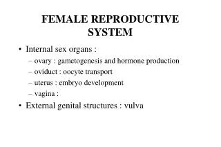
The human Female Reproductive System The human Female Reproductive System is almost entirely hidden within the pelvis. It consists of organs that enable a woman to produce eggs (ova), to have sexual intercourse, to nourish and house the fertilized egg (ovum) until it is fully developed, and to give birth
Female Reproductive Organs internalreproductive organs - Ovaries - Uterine tubes - Vagina Externalreproductiveorgans -- mons pubis -- labia majora -- labia minora -- clitoris and -- vestibular glands
Ovaries These are paired structures. - almond shaped, greyish pink in color, - 3 cm long. 1.5 cm wide and 1 cm thick. - placed on each side of the uterus.. Ovary is attached to the posterior surface of the inner body wall by mesovarium. Ovaries are further supported by suspensory and ovarian ligaments.
Ovarian structure Ovary is divisible into 2 regions: - Cortex ( contains ovarian follicles ) - Medulla( receives blood vessels and nerves at the hilum ) Surface of the Ovary covered by a cuboidal cell layer of Ovarian surface epithelium. Beneath the epithelium, the ovary is surrounded by a tough coat tunica albuginea. It is made up of collagenous tissue. After puberty the cortex forms the major part of the ovary. Cortex is filled with stroma composed of collagen. Ovarian follicles are embedded in the stroma.
Ovarian Follicles The Ovary of foetus at 5 months gestation has 7 million Oocytes. At birth the ovary of the child contains about 1 million Oocytes. Due to further degeneration, At the time of Puberty only about 40000 Oocytes remain. Of the 40,000 oocytes only about 400 undergo ovulation during the reproductive years.
Ovarian Follicles At birth, the primordial follicles are found in the superficial zone of the cortex. They contain primary oocytes (about 25mm in diameter). Each one of them is surrounded by a single layer of flat follicular cells. The various follicular stages are: -- Primary follicles -- Secondary follicles -- Tertiary follicles
Ovarian Follicles Primary Follicle – The follicle cells are converted from squamous to cuboidal cells. Follicular membrane or membrana granulosa becomes multilayered. The Oocyte increases in size. It has an outer thick layer called zona pellucida. The follicular cells divide and form granulosa cells. Secondary Follicle – It is about 20m thick. The granulosa cells surround the oocyte and form a mound of cells called the cumulus ovaricus. The inner and outer theca become prominent. Theca interna is well established. Tertiary Follicle – Only one follicle reaches tertiary stage. It increases in size (2 mm diameter), Now it is called the graffian follicle. The oocyte and the corona radiata break away and float freely in the follicular fluid Finally the wall of the follicle ruptures and the contents are released into the peritonium.
CORPUS LUTEUM It is formed after ovulation. The walls of empty follicle collapses and fold extensively. The granulosa cells of the theca externa get enlarged. They are now termed as Luteal cells. In pregnancy Corpus luteum persists. Other wise, it degenerates after 10-12 days. The connective tissue cells get enlarged. It becomes white in color and is now called corpus albicans. In course of time the corpus albicans shrinks and disappears.
UTERINE TUBES(Fallopian Tubes) There are 2 Uterine tubes or fallopian tubes (Oviducts) connect the uterus withthe ovaries. Each tube is about 10 cm length. The terminal part of the tube is enlarged to form theinfundibulum. It opens in the peritonial cavity. The opening is called Ostia.
UTERINE TUBES(Fallopian Tubes) The Uterine tube consists of 3 parts: - ampulla( nearer to the infundibulum, longest part) - isthmus( nearer to the uterus, narrower) - uterine(tubular part entering in the uterus)
UTERUS -- pear shaped, -- a hollow thick walled muscular organ. -- about 7.5 cm long and 5 cm wide. -- weighs about 50 gm. -- during pregnancy it may go upto 1 Kg. -- houses the foetus during pregnancy.
Parts of the Uterus: Parts of the Uterus: -- the large rounded part is fundus -- the middle part is body. -- the narrower part is cervix which directs inferiorly. Uterus continues as the cervical canal and opens into the vagina through the Ostium.
Wall of the Uterus: the outermost layer is the perimetrium or Serous layer. the next layer myometrium or muscular coat is the major part of the wall. the innermost functional layer is endometrium or mucous membrane which undergoes menstrual changes and sloughing during female sex cycle.
VAGINA It extends from uterus to outside. It is a fibro-muscular tube. It is a copulatory organ. It is 10 cm. long. This vaginal passage used during intercourse and allows menstrual flow and child birth.
External Genitalia The External genitalia is known as the Vulva or pudendum. It consists of the Vestibule and its surrounding structures. The vestibular region remains in between the 2 Labia majora. It contains the Vaginal opening and the Urethral Opening. The vestibular region is surrounded by the Mons pubis anteriorly and Labia majora and Labia minora on the lateral sides.
External Genitalia Mons pubis - is a rounded eminence situated anteriorly. - made up of sub-cutaneous adipose tissue. - covered by coarse hair at the tiem of puberty. - corresponds to similar structure in the female. Labia Majora - are longitudinal folds of skin. - form the outer boundary for the vestibule. Labia Minora - are 2 small skinfolds lie between the labia majora. - remain nearer to the vaginal opening. Clitoris - is an erectile structure. - found in the anterior margin of the vestibule. - is a sensitive organ having many receptors. - is homologous with male penis.
External Genitalia Hymen Vaginae– is a thin mucous membrane. - is found within the vaginal orifice or opening. - has no established function. In young women it may normally get torn during physical exercise. It should be removed to allow menstrual flow. In some women it may be absent. External Urethral Opening: -- remains as a small cleft –- is about 2.5 cm below the clitoris -- is anterior to the Vaginal opening