Comprehensive Sialic Acid Standards for Glycosylation Analysis
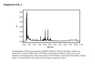
Explore the characterization of sialic acid in Dsp N-glycosylations using fluorescent chromatograms from SE-HPLC and RP-HPLC separations. The article also includes a table showing quantitative determination of sialic acid moieties in different fractions.

Comprehensive Sialic Acid Standards for Glycosylation Analysis
E N D
Presentation Transcript
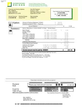
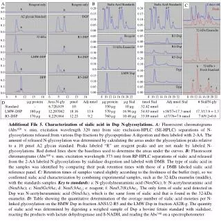
A Reagent only Reagent only B Sialic Acid Standards Sialic Acid Standards 2 days old C .05 e e b b d d R 0.20 0.20 a a R Fresh 0.0 0.15 0.15 A2 glycan Standard DGP R c c 0.2 0.10 0.10 f f 0.1 0.05 0.05 R 32-kDa Enamelin 0.0 0.00 0.00 32-kDa R3-DSP Reagent R3-Dsp 0.05 0.05 0.2 0.00 0.00 0.1 R R4-Dsp 0.05 0.40 32-kDa Enamelin R 0.0 0.00 0.30 LMW-DSP R4-DSP R5-Dsp 0.2 0.20 0.20 0.10 0.1 0.10 32-kDa + Fresh Standard b R R 0.0 Dgp 0.0 0.10 LMW Dsp R5-DSP DPP 0.15 0.05 0.2 d a 0.10 0.00 0.1 c e 0.05 Dpp 0.05 R R f 0.0 0.00 0.00 6 6 9 9 12 12 15 15 18 18 8 10 12 14 16 18 20 22 8 10 12 14 16 18 20 22 8 10 12 14 16 18 20 22 µg protein Area N-gly pmol Adj nmol µg protein µg Sial nmol Sial Adj nmol Sial # Sial/N-gly D Standard - 6,720,819 10 - 350 µg 10 µg 32.42 nmol - - LMW-DSP 180 µg 12,207042 18.16 13.6 570 µg 16.96 µg 54.83 nmol x18/57=17.3 nmol 17.3/13.6 = 1.3 R5-DSP 170 µg 8,229,864 12.25 9.2 760 µg 10.48 µg 33.89 nmol x17/76=7.6 nmol 7.6/9.2=0.8 Additional File 5. Characterization of sialic acid in Dsp N-glycosylations. A:Fluorescent chromatograms (Abs420 v. min; excitation wavelength 320 nm) from size exclusion-HPLC (SE-HPLC) separations of N-glycosylations released from various Dsp fractions by glycopeptidase A digestion and then labeled with 2-AA. The amount of released N-glycosylation was determined by calculating the areas under the glycosylation peaks relative to a 10 pmol A2 glycan standard. Peaks labeled “R” are reagent peaks and are not made by labeled N-glycosylations. Red dotted lines show the baselines used to determine the areas under the curves. B: Fluorescent chromatograms (Abs448 v. min; excitation wavelength 373 nm) from RP-HPLC separations of sialic acid released from the 2-AA labeled N-glycosylations by sialidase disgestion and labeled with DMB. The type of sialic acid in the samples was identified by comparing their peak retention times with those of a DMB labeled sialic acid reference panel. C: Retention times of samples varied slightly according to the freshness of the buffer (top), so we confirmed sialic acid characterization by combining experimental samples, such as the 32-kDa enamelin (middle), with the standards samples. Key to standards: a: N-glycolylneuraminic acid (Neu5Gc); b: N-acetylneuraminic acid (Neu5Ac); c: Neu5Gc9Ac; d: Neu5,9Ac2; e: reagent; f: Neu5,7(8),9Ac3. The only form of sialic acid detected in Dsp was N-acetylneuraminic acid (Neu5Ac), which is the same form of sialic acid that is found in the 32-kDa enamelin. D: Table showing the quantitative determination of the average number of sialic acid moieties per N-linked glycosylation on the HMW Dsp in fraction ANS1/2-R5 and the LMW Dsp in fraction AS2R-g. The quantity of sialic acid was determined by digesting a weighed sample of Dsp a bovine fetuin standard with sialidase, reacting the products with lactate dehydrogenase and ß-NADH, and reading the Abs340 on a spectrophotometer.