Download
1 / 40
400 likes | 428 Vues
Annotate diagrams of male and female reproductive systems to show structures and functions. Understand male and female reproductive system anatomy and hormone functions. Learn about the menstrual cycle, ovulation, and in vitro fertilization (IVF). Explore the roles of testosterone, estrogen, and progesterone in reproduction. Discuss ethics in IVF and William Harvey's research on sexual reproduction. Enhance your biology knowledge with detailed annotations and animations.

E N D
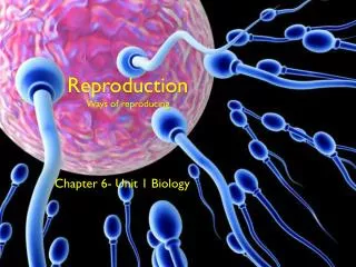
Skill: Annotate diagrams of the male and female reproductive system to show names of structures and their functions.
Male Reproductive System: Structure and Function: • Testes~ male gonads (produce sperm and hormones • Seminiferous tubules~ sperm formation • Leydig cells~ secrete testosterone • Epididymis~ sperm develop motility and mature • Scrotum~ helps regulate temp. • Vas deferens (sperm duct)~ sperm propelled by peristalsis • Exocrine glands: • Seminal vesicle~ adds fluids and fructose to sperm. • Prostate gland~ adds anticoagulant; nutrients, more fluids • Bulbourethral glands~ acid neutralizer • Penis/urethra~ semen delivery
• Skill: Annotate diagrams of the male and female reproductive system to show names of structures and their functions.
Testosterone • A gene on the Y chromosome causes embryonic gonads to develop as testes and secrete testosterone. • Roles of testosterone • Pre-natal development of male genitalia • Sperm production • Development of secondary sex characteristics
Female Reproductive System: Structure and Function Be able to annotate • The Human Female • Ovaries~ female gonads– make eggs • Follicle~ one egg surrounded by one or more layers of follicle cells that nourish and protect egg cell • Corpus luteum~ post-ovulation tissue of follicle– secretes hormones (estrogen and progesterone) • Oviducts/Fallopian tubes~ where fertilization occurs • Uterus/endometrium~ womb/lining • Cervix/vagina~ sperm receptacle
Female hormones • Estrogen and progesterone cause pre-natal development of female reproductive organs and female secondary sexual characteristics during puberty. • The menstrual cycle is controlled by negative and positive feedback mechanisms involving ovarian and pituitary hormones.
Ovulation and the menstrual cycle • Really cool 3d ovulation animation video: http://catalog.nucleusinc.com/generateexhibit.php?ID=67312&ExhibitKeywordsRaw=&TL=&A=2
The Menstrual Cycle The roles of FSH, LH, estrogen and progesterone in the menstrual cycle are expected. • Estrous cycles/estrus (many mammals) “heat”, endometrium reabsorbed by body. • Menstrual cycle: involves menstruation: shedding of endometrium. • Menstrual cycle~ (Know the hormones!) • FSH causes: • follicle growth • estrogen secretion • Estrogen: • Repairs uterine lining after menstruation • Stimulates LH secretion (when levels peak)
LH (luteinizing hormone) causes: • Ovulation • less estrogen • More progesterone • Follicle to develop into corpus luteum (continues releasing estrogen and progesterone.) • Progesterone: maintains uterine lining (endometrium) to prepare for implantation. • If no implantation, estrogen and progesterone levels fall, causing menstruation and cycle begins again.
Not in course guide anymore: Annotate a graph showing hormone levels in the menstrual cycle, illustrating the relationship between hormone levels and ovulation, menstruation, and thickening of the endometrium
Menstrual Cycle Menstrual Cycle Animation http://www.pbs.org/wgbh/amex/pill/sfeature/sf_cycle.swf Another http://www.sumanasinc.com/webcontent/animations/content/ovarianuterine.html
In Vitro Fertilization (IVF) • Drug injections stop menstrual cycle • Large doses of FSH stimulate development of many follicles (rather than one as in normal cycle) Superovulation… • HCG is injected to mature the oocytes in the follicles and loosen them so they can be retrieved. • Oocytes are retrieved (an invasive procedure) • Collect and Prepare the Sperm – the healthiest ones are concentrated during preparation. • Inseminate Eggs • Incubate fertilized eggs • Transfer successful Embryos to the Uterus with a long plastic tube • Pregnancy test for implantation • http://www.sumanasinc.com/webcontent/animations/content/invitrofertilization.html • Discuss the Ethical Issues associated with IVF!
Application: William Harvey’s investigation of sexual reproduction in deer. • William Harvey failed to solve the mystery of sexual reproduction because effective microscopes were not available when he was working, so fusion of gametes and subsequent embryo development remained undiscovered. • Debunking the ’Soil and Seed’ Theory • William Harvey studied the sexual organs of female deer after mating in an effort to identify the developing embryo • He was unable to detect a growing embryo until approximately 6 – 7 weeks after mating had occurred • He concluded that Aristotle’s theory was incorrect and that menstrual blood did not contribute to the development of a fetus • Harvey was unable to identify the correct mechanism of sexual reproduction and incorrectly asserted that the fetus did not develop from a mixture of male and female ‘seeds’
Annotate a light micrograph to show the location and function of: interstitial cells (Leydig cells) –produce testosterone germinal epithelium cells constantly divide by mitosis making diploid cells developing spermatozoa –become viable sperm Sertoli cells.– nurse cells (provide nutrients to developing sperm)
Annotate a light micrograph to show the location and function of: • interstitial cells (Leydig cells) • germinal epithelium cells • developing spermatozoa • Sertoli cells. • Note: Developing sperm move toward the lumen (center) of the seminiferous tubule
Spermatogenesis Constant from puberty until death! Germinal epithelium cells (Primordial germ cells) (2N)~ differentiate into…. Spermatogonia (2N)~ stem cells that give rise to sperm Repeated mitosis into…. Primary spermatocyte (2N) Meiosis I produces… Secondary spermatocytes (1N) Meiosis II produces… Spermatids (1N) not fully developed. Development helped by Sertoli cells Sperm cells (1N) http://highered.mcgraw-hill.com/olc/dl/120112/anim0043.swf
The Ovary Annotate to show the location and function of: Germinal epithelium Primary follicles Mature follicle Secondary oocyte
Oogenesis in ovary Germinal epithelium (Primordial germ cells) (2N)– Undergo Mitosis and differentiation to make… More diploid cells (oogonia) which develop into… Primary oocytes (2N) Stopped at prophase I of meiosis I Surrounded by a layer of follicle cells. Primary oocyte + follicle cells = primary follicle All 400,000 primary follicles formed before birth
Between birth & puberty; egg cells and follicles grow Every menstrual cycle, a few follicles develop. Here’s how: FSH (follicle stimulating hormone) causes growth of follicles; Primary oocyte completes meiosis I forming two haploid cells: A small polar body (dies) Secondary oocyte – 1N (undergoes prophase II of meiosis then stops.) Follicle ruptures (ovulation) and releases secondary oocyte into oviduct. After fertilization, Meiosis II is completed. This forms 2 cells: Ovum (the egg, already fertilized) 2nd polar body (dies) Note: Polar bodies degenerate
Annotate: a mature egg Note: The Zona pellucida is composed of glycoproteins. With the cortical granules they will be involved in the acrosome reaction at fertilisation
Annotate: a mature sperm Acrosome
Compare Spermatogenesis to Oogenesis http://wps.aw.com/bc_martini_eap_4/40/10469/2680298.cw/content/index.html
Fertilization Arrival of sperm at egg Sperm break through layers of follicle cells Acrosome reaction~ acrosome releases hydrolytic enzymes which digest the zona pellucida, allowing the sperm in. Fusion: Egg and sperm plasma membranes fuse and sperm nucleus enters egg. Fast block to polyspermy~ membrane depolarization prevents multiple fertilizations…. Cortical reaction~ enzymes from cortical granules make zona pellucida harden and creates a... Slow block to polyspermy (created by development of fertilization membrane). A zygote has been produced. C:\Documents and Settings\BBAUGHMAN\Desktop\bio powerpoints\Chapter 38 BDOL IC
Early embryo development Cell division (Mitosis) continues until a blastocyst (hollow ball of cells) is formed. The blastocyst will implant in uterus. Video: http://www.pbs.org/wgbh/nova/miracle/program.html Taking Shape
Pregnancy Gestation~ pregnancy Begins at implantation of blastocyst in endometrium of uterus. 1st trimester:(1st 3 months) Main period of organogenesis fetus (week 8) HCG (human chorionic gonadotropin) hormone– secreted by embryo– maintains progesterone secretion by ovary (during early pregnancy)
The placenta and umbilical cord Placenta– organ with embryonic and maternal blood vessels (nutrient, gas & waste diffusion) Placental villi- contain fetal capillaries Intervillus spaces– where maternal blood flows NOTE: The placenta secretes estrogen and progesterone to maintain the endometrium Umbilical cord Umbilical vein carries oxygenated, nutrient-rich blood to baby Umbilical arteries carry wastes and CO2 away from baby http://www.pennmedicine.org/encyclopedia/em_DisplayAnimation.aspx?gcid=000101&ptid=17 http://catalog.nucleusinc.com/generateexhibit.php?ID=69893&ExhibitKeywordsRaw=&TL=&A=2
http://www.childrenshospital.org/az/Site636/mainpageS636P0.htmlhttp://www.childrenshospital.org/az/Site636/mainpageS636P0.html
Amniotic Sac and amniotic fluid Supports and protects fetus
Birth End of pregnancy signalled by drop in progesterone Oxytocin is produced, causing Labor~uterine contractions Positive feedback increases oxytocin production and contractions. • Note: Estrogen increases oxytocin receptors in uterus
Animal Reproduction comparisons Application: The average 38-week pregnancy in humans can be positioned on a graph showing the correlation between animal size and the development of the young at birth for other mammals. Spectrum ranging from Altricial species: newborns are helpless to Precocial species: newborns are mobile, aware etc. Fertilization in animals can be internal or external.
Nature of science: Assessing risks and benefits associated with scientific research—the risks to human male fertility were not adequately assessed before steroids related to progesterone and estrogen were released into the environment as a result of the use of the female contraceptive pill. (4.8)
Ultrasound http://health.howstuffworks.com/adam-200128.htm
Outline the origin and the role of the follicle stimulating hormone (FSH), testosterone, and lutenizing hormone (LH) in spermatogenesis. Follicle Stimulating Hormone: stimulates primary spermatocytes to undergo the first division of meiosis. Lutenizing Hormone: stimulates secretion of testosterone by testes Testosterone: stimulates development of secondary spermatocytes into mature sperm. Origin: FSH and LH =Pituitary Testosterone = testes
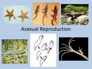
Odd Reproductive cycles • Parthenogenesis unfertilized egg development; haploid, sterile adults (honeybees) • Hermaphroditismboth male & female reproductive systems; sessile & burrowing organisms (earthworms) • Sequential hermaphroditism reversal of gender during lifetime•protogynous(female 1st) •protandrous(male 1st)