ACCIDENTAL HYPOTHERMIA
ACCIDENTAL HYPOTHERMIA. Melanie Stander. Defined as decline in core body temperature below 35C. Mild : 35-32 C Moderate : <32 – 28 C Severe : <28 C. Risk Factors. Extremes of age Ethanol abuse Malnutrition Poverty Mental disease Neuroleptic drugs Hypothyroidism Cold-water immersion

ACCIDENTAL HYPOTHERMIA
E N D
Presentation Transcript
ACCIDENTAL HYPOTHERMIA Melanie Stander
Defined as decline in core body temperature below 35C. • Mild : 35-32 C • Moderate : <32 – 28 C • Severe : <28 C
Risk Factors • Extremes of age • Ethanol abuse • Malnutrition • Poverty • Mental disease • Neuroleptic drugs • Hypothyroidism • Cold-water immersion • Winter sports athletes
Mortality & Prognosis • Despite immediate treatment, improved prehospital survival and rewarming techniques in hospital, accidental hypothermia is associated with a high mortality ranging from 30-80%. • Walpoth, 1990, 11 pts – 35% mortality • Kornberg, 1996, 24 pts – 87% mortality • Hauty, 1987, 82% mortality, no survivors with temps <20 C.
Farstad, 2001, 26 pts – 79% mortality • Gilbert, 2000, successful resus of pt with central temp of 13.7 C. • K levels >10, pH<6.5 & low PaO2 are poor prognostic indicators. • Prognosis worsens in pts with asphyxia
Case report from dept of Cardiothoracic surgery, University Hospital Ostrava, Czech Republic – successful resus of 26 yr old male, found 15 hours after a suicide attempt (carbamazepine OD) in deep hypothermia of 19 C with circulatory arrest. ECC was used to rewarm pt – pt showed no neurological deficit at time of discharge from hospital.
Physiology • Thermoregulation is coordinated in pre-optic anterior hypothalamus • 4 mechanisms of heat loss: 1. Radiation (55%) 2. Evaporation (airway-5%, skin-25%) 3. Conduction (10%) 4. Convection (5%)
Predisposition • Decreased heat production. • Increased heat loss. • Impaired thermoregulation.
Effects on Organ Systems • CVS • CNS • Renal system • Respiratory system • Haematological changes • Acid-base • Endocrine
Cardiovascular system • Initial tachycardia, then bradycardia. • Pulse decreases by 50% at 28 C. • Bradycardia is due to decreased spontaneous depolarization of pacemaker cells (refractory to Atropine). • Cardiac index and MAP decreased • J waves (Tomaszewkski, 1938)
J waves are potentially diagnostic but not prognostic. • Appear at temps below 32 C. • Size of J wave does increase with temp decrease. • Normally upright in aVL, aVF and L praecordial leads. • May be result of hypothermic ion flux alterations, with delayed depol or repol of LV wall.
All atrial and ventricular dysrrhythmias are common. • As hypothermia worsens, first the PR interval, then QRS interval and finally the QT interval becomes prolonged. • Sinus rhythm and junctional rhythms are common. • AF often occurs at temps below 32 C. This usually converts spontaneously during rewarming but mesenteric emboli are a hazard.
Asystole and VF occur spontaneously at core temps below 25 C. • Decrease in transmembrane RP occurs which decreases the ventricular dysrrhythmia threshold. • A cold heart has a large dispersion of repolarization which facilitates the development of a conduction delay. • The AP is also prolonged.
The CNS • Cerebral depression • 6-7% decrease in metabolism for each 1 C decline in temperature.
Renal System • Cold diuresis regardless of state of hydration. • Temps of 27-30 C : only 50% renal blood flow. • Cold diuresis is GF which doesn’t clear nitrogenous waste products. • Initial relative central hypervolaemia due to peripheral vasoconstriction, cold diuresis helps to decrease the vasoconstriction-induced capacitance vessel overload.
Oxidative tubular activity depressed – decreased Na and water resorption. • Can render pt hypovolaemic.
Respiratory System • Initial stimulation of respiration. • Progressive decrease in resp. minute volume which is proportional to the drop in metabolism. • CO2 production decreases. • Also CO2 retention and respiratory acidosis can occur. • Viscous brochorrhea, decreased ciliary motility, noncardiogenic pulmonary oedema. • Oxy-Hb curve shifted to left.
Haematological Changes • Coagulation cascade impaired. • Plasma fibrinolytic activity is enhanced. • Platelets are sequestered and function poorly. • Volume depletion – haemoconcentration – increased blood viscosity – thrombosis.
Acid-Base Balance • Lactic acidosis due to decreased peripheral perfusion with compensatory respiratory alkalosis. • Respiratory depression – respiratory acidosis.
Endocrine System • Initial hyperglycaemia (inhibition of insulin release and decreased use of insulin peripherally). • ACTH and TSH increased. • Elevated catecholamines and cortisol.
Diagnosis • Rectal or oesophageal core temperature measurement using a low-reading thermometer. • Wide-ranging clinical presentation. • Reflexes brisk down to 32 C then decrease and then disappear at around 26 C. • The knee jerk, when present, is the last reflex to disappear and the first to reappear with warming.
Investigations • FBC, U&E, Glucose, ABG, B/Cs and consider TFTs. • The Hct is high due to decreased plasma volume. • Normal WCC doesn’t exclude infection. • Hypothermia enhances the cardiac toxicity and obscures ECG changes of hyperkalaemia. • Hypokalaemia is most common with chronic hypothermia.
Serum enzymes are elevated – rhabdomyolysis is commonly associated with cold exposure. • Leucopaenia and thrombocytopaenia usually reverse with rewarming. • ECG • X-rays and other investigations according to clinical scenario.
ABGs – blood gas analysers warm blood to 37 C which increases the partial pressure of dissolved gases. ABG shows higher O2 and CO2 levels and lower pH than pt’s actual values. Current practise – correction of ABGs for temp is unnecessary as a guide for therapy.
Management • Prehospital • ED - Initial - Volume resus - ALS - REWARMING
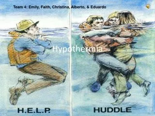
Prehospital • Prevent heat loss and rewarm the core before the shell. • Rescue, examine, insulate & rapidly transport. • A, B, C. • Remember to feel much longer for a pulse (ACLS guidelines 30-45 seconds). • Handle the pt gently – a cold myocardium is very susceptible to VF.
Defib attempts are usually unsuccessful until the core temp is above 28 C. • Keep the pt dry and insulated. • Give a fluid challenge. • Avoid muscular exertion • Inhalation of heated and humidified air.
Airway Rewarming • Heat is absorbed in the nasopharyngeal region & transferred the short distance to the brainstem where thermoregulatory, ventilatory & circulatory control centres are located. • Increasing the temp of these centres maintains & restores the drive for shivering, ventilation & cardiac output, facilitating rewarming by endogenous heat production. • Nearby arteries are warmed (eg. Basilar) that lead to the circle of Willis.
ED • Continuous core temperature monitoring. • Rectal temperature is most practical. • Tympanic temperature is most closest to hypothalamic temp. • Again: A, B, Cs. • NGT, urinary catheter, cardiac monitoring.
Rapid volume expansion is critical. • Initial heated fluid (40 –42 C) bolus of 250-500ml 5% dextrose in normal saline (don’t give R/L as the cold liver ineffectively metabolises lactate.) • Microwave 1L of crystalloid for 2 mins on high – shake before administration to avoid hot spots. • Decrease length of IV tubing.
CPR – chest wall elasticity and pulmonary compliance are decreased with cold so more force is needed to depress the chest wall. • Concern that in the severely hypothermic pt, cardioactive medications can accumulate to toxic levels in peripheral circulation if given repeatedly. Withhold IV drugs if core temp <30 C. If temp >30 C administer IV meds with increased intervals between doses.
Rewarming • Passive vs. Active • Active : external or internal
Passive External Warming • No added heat. • Non invasive. • Treatment of choice for mild hypothermia. • The pt must be able to metabolically generate sufficient heat to maintain an acceptable rate of spontaneous rewarming.
Keep ambient temp >25 C. • Keep head covered – 30% of body heat can be lost via the head. • Rewarming rates vary between 0.5-2.0 C/hr.
Indications for Active Rewarming • CVS instability • Moderate or severe hypothermia (<32 C). • Inadequate rate or failure to rewarm. • Endocrine insufficiency • Traumatic or toxological peripheral vasodilation. • Secondary hypothermia impairing thermoregulation.
Active rewarming involves the direct transfer of exogenous heat to the pt.
Active External Rewarming • Heat sources – immersion in warm water, hot water bottles and heaters, Bair-hugger. Beware of thermal injury to vasoconstricted hypoperfused skin. • “Afterdrop” –peripheral vasodilation and shunting of cold, acidotic blood full of metabolic waste to core tissues.
Active Internal Rewarming 1. Humidified, heated oxygen Via face mask or ETT. Inspired air should not exceed 40 C. Avoidance of afterdrop. Maintenance of adequate oxygenation is vital (shift of oxyHb dissociation curve).
2. Warmed IV fluids 3. Peritoneal Lavage Infuse 1L of warmed balanced salt solution for one min and then drain. Fluid rates of 10-12L/hr are possible. Rosen’s suggests 2L infused and retained for 20-30 mins and then aspirated. Hepatic rewarming reactivates depressed detoxification and enzymes. Exacerbates hypokalaemia.
4. Pleural Lavage Preferably left side. Infuse 1L warmed N/S for 1 min and then drain. Can use one tube anteriorly in 2nd or 3rd ICS MCL and second tube in PAL at 5th to 6th ICS.
5. Diathermy Restores body heat by ultrasonic waves, microwaves or shortwaves. Involves the conversion of energy waves into heat. Noninvasive way to deliver heat to core tissues.
6. Extracorporeal Blood Warming CPB, AV rewarming, VV rewarming & haemodialysis. CPB: major advantage is preservation of flow if mechanical cardiac activity is lost during rewarming. CAVR: blood pressure must be at least 60mmHg. Ipsilateral femoral artery and contralateral femoral vein.
VVR: blood removed from CVP, heated to 40 C and returned via second CVP or large peripheral venous catheter. Haemodialysis: portable and efficient. Consider in electrolyte abns, RF or intoxication with a dialysable substance.
Septicaemia and Hypothermia • Host defences are compromised and there is a predisposition to infection. • Usual signs of infection including fever are absent. • Decreased BM release and circulation of neutrophils. Impaired neutrophil migration and bacterial phagocytosis. • Role of antibiotic prophylaxis in adults is not clear. • Elderly patients with thermoregulatory failure have a high mortality and should be considered to be septic.
References • Radim Brát et al, Rewarming from severe accidental hypothermia with circulatory arrest, Biomedical papers 148(1), 51-53(2004). • Accidental Hypothermia, Chamonix, 1998. • Dr John Hayward, On the effectiveness of airway heat donation. • Circulation 2005;112;136-138;published online Nov 28,2005.
5. State of Alaska Cold Injuries Guidelines, Alaska Multi-level, 2003 version. Prepared by Dept of Health & Social Services, Division of Public Health, Section of Community Health and EMS. 6. Robert Douwens, Hypothermia Recognition and Treatment. 7. Rosen’s Emergency Medicine, Concepts and Clinical Practice, 5th edition, volume 3. 8. Oxford Handbook of Trauma for Southern Africa, Nichol & Steyn 2004.