The Cell Cycle
The Cell Cycle. Imagine your skin…. Cells have a life cycle. It goes from birth…to birth…to birth…to birth… Is there death? Eventually… Cells (most human cells at least) are limited in the number of times they can reproduce.

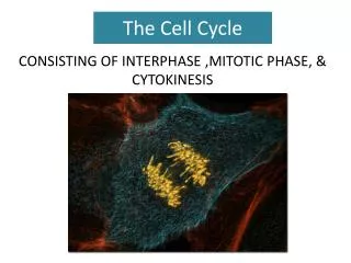
The Cell Cycle
E N D
Presentation Transcript
Cells have a life cycle • It goes from birth…to birth…to birth…to birth… • Is there death? • Eventually… • Cells (most human cells at least) are limited in the number of times they can reproduce. • Infant skin cells placed in a Petri dish may reproduce 100 or more times • A 60 year old’s skin cells may only reproduce about 20 times.
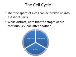
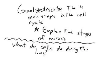
So what’s in between each “birth” event? • Life. • The cell carries out it’s life functions. It does its job. • The life cycle of a cell… from birth to life to birth is called the CELL CYCLE.
By Convention… • We normally talk about INTERPHASE as the stage that marks the “beginning of the cell cycle.
INTERPHASE • The stage of a cell’s life in which the cell is doing one of two things: • Living out its life and doing its job • Preparing for cell division – the “birth” of two new cells
Interphase Time to get ready to make new cells! • 90% of cell life cycle • cell doing its “everyday job” • prepares for duplication if triggered I’m working here! Time to make new Cells!
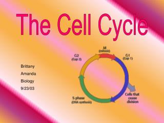
Cell cycle • Cell has a “life cycle” cell is formed from a mitotic division cell grows & matures to divide again cell grows & matures to never divide again G1, S, G2, M liver cells G1G0 epithelial cells,blood cells, stem cells brain / nerve cells muscle cells
G0 signal to divide Interphase • Divided into 3 phases: • G1= 1stGap (Growth) • cell doing its “everyday job” • cell grows • S= DNA Synthesis • copies chromosomes • G2 = 2ndGap (Growth) • prepares for division • cell grows (more) • produces organelles,proteins, membranes
Understanding Chromosome Structure and Terminology • During G1
Understanding Chromosome Structure and Terminology • During S
Understanding Chromosome Structure and Terminology • During G2
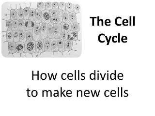
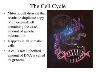
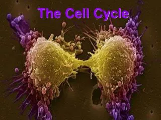
MITOSIS • NOW we are ready to “give birth” to new cells! • We have replicated the genetic material • We now need to “divvy up” one complete set of genes to each cell that will be “born” • THIS is mitosis – the division of the nucleus
Mitosis • Dividing cell’s DNA (chromosomes) between 2 daughter nuclei • 4 phases • prophase • metaphase • anaphase • telophase
Why do cells divide by MITOSIS? • For reproduction • asexual reproduction • one-celled organisms • For growth • from fertilized egg to multi-celled organism • For repair & renewal • replace cells that die from normal wear & tear or from injury GOAL of MITOSIS = DAUGHTER CELLS that are IDENTICAL to the PARENT CELLS!! amoeba CLONES!!!
Cytoskeleton Fibers • Fibers that support the cell as well as move things around in the cell • VERY IMPORTANT in the events of MITOSIS • THEY MOVE THE CHROMOSOMES! • THEY CUT THE CELL IN TWO!
Cytoskeletal Fibers • Made of PROTEIN • 3 Types • Intermediate Filaments • Microfilaments • Microtubules
Intermediate filaments • No real role in mitosis • More permanent • Made of Keratin family of proteins • Support • Fix position of organelles • When shape is important
Microfilaments • Solid rods • Made of actin protein • Bear tension (pulling forces) • Support and change cell shape – 3D network just inside cell membrane • Responsible for Cleavage furrow in cell division (actin interacting w/ myosin – much like in muscle cell)
Microtubules • Straight hollow rods • Made of tubulin – a protein • Microtubules elongate by adding tubulin molecules to the ends of the tube • Can be disassembled and reassembled elsewhere • Provide: • Shape; support • “tracks” • guide secretory vesicles to plasma membrane • Separation of chromosomes during cell division
Microtubules and Cell Division • Centrosomes • Centrioles • May help organize spindle • Animal cells only • Spindle Fibers
I.P.M.A.T. Overview of the Cell Cycle interphase prophase (pro-metaphase) cytokinesis metaphase anaphase telophase
green = key features Prophase • Chromatin condenses • visible chromosomes • chromatids • Centrioles move to opposite poles of cell • animal cell • Protein fibers cross cell to form mitotic spindle • microtubules • actin, myosin • coordinates movement of chromosomes • Nucleolus disappears • Nuclear membrane breaks down
green = key features Transition to Metaphase • Prometaphase • spindle fibersattach to centromeres • creating kinetochores • microtubules attach at kinetochores • connect centromeres to centrioles • chromosomes begin moving
Kinetochore • Each chromatid has own kinetochore proteins • microtubules attach to kinetochore proteins
green = key features Metaphase • Chromosomes align along middle of cell • metaphase plate • meta = middle • spindle fibers coordinate movement • helps to ensure chromosomes separate properly • so each new nucleus receives only 1 copy of each chromosome
green = key features Anaphase • Sister chromatids separate at kinetochores • move to opposite poles • pulled at centromeres • pulled by motor proteins “walking”along microtubules • actin, myosin • increased production of ATP by mitochondria • Poles move farther apart • polar microtubules lengthen
Separation of chromatids • In anaphase, proteins holding together sister chromatids are inactivated • separate to become individual chromosomes 1 chromosome 2 chromatids 2 chromosomes single-stranded double-stranded
green = key features Telophase • Chromosomes arrive at opposite poles • daughter nuclei form • nucleoli form • chromosomes disperse • no longer visible under light microscope • Spindle fibers disperse • Cytokinesis begins • cell division
Cytokinesis in Plants • Plants • cell plate forms • vesicles line up at equator • derived from Golgi • vesicles fuse to form 2 cell membranes • new cell wall laid down between membranes • new cell wall fuses with existing cell wall
Origin of replication chromosome: double-stranded DNA replication of DNA elongation of cell ring of proteins cell pinches in two Evolution of mitosis • Mitosis in eukaryotes likely evolved from binary fission in bacteria • single circular chromosome • no membrane-bound organelles
prokaryotes (bacteria) Evolution of mitosis • A possible progression of mechanisms intermediate between binary fission & mitosis seen in modern organisms protistsdinoflagellates protistsdiatoms eukaryotes yeast eukaryotes animals