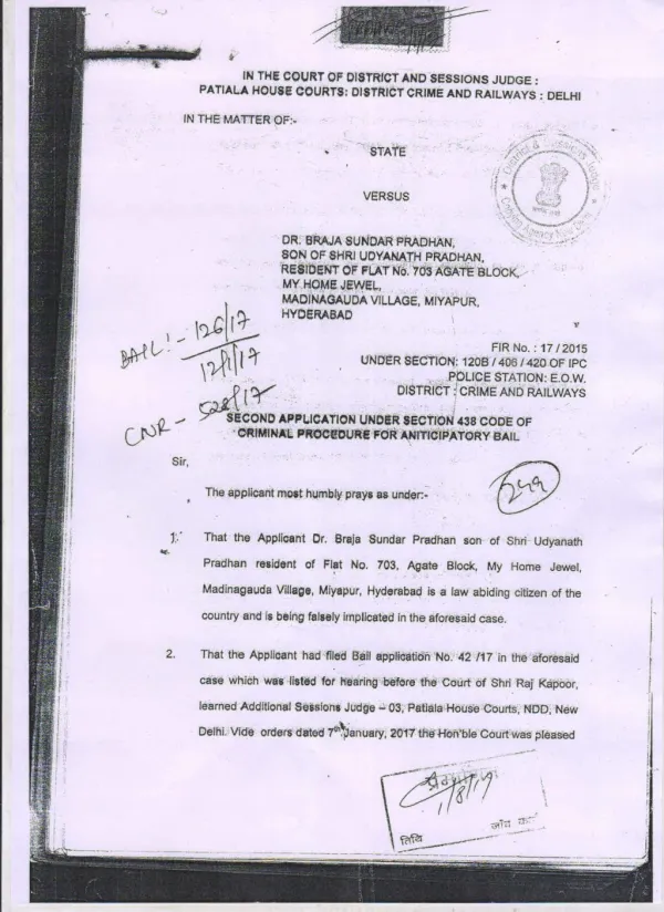
Satish Pradhan Dnyanasadhana College, Thane
Satish Pradhan Dnyanasadhana College, Thane Department of Chemistry T.Y.B.Sc. Analytical Chemistry Paper-IV Sem-V UV-Visible spectroscopy. Contents. 4.2 Molecular Spectroscopy – III (04 L) 4.2.1 Recapitulation of basic concepts

Satish Pradhan Dnyanasadhana College, Thane
E N D
Presentation Transcript
Satish Pradhan Dnyanasadhana College, Thane Department of ChemistryT.Y.B.Sc. Analytical ChemistryPaper-IV Sem-VUV-Visible spectroscopy
Contents • 4.2 Molecular Spectroscopy – III (04 L) • 4.2.1 Recapitulation of basic concepts • 4.2.2 Instrumentation, Principle and working of single and double beam spectrophotometers. • 4.2.3 Applications of UV- Visible Spectrophotometery
4.2.1 Recapitulation of basic concepts • What is Electromagnetic Radiation • Electromagnetic radiation, or light, is a form of energy whose behavior is described by the properties of both waves and particles. The optical properties of electromagnetic radiation, such as diffraction, are explained best by describing light as a wave.
Properties of electromagnetic radiation • Frequency: The number of oscillations of an electromagnetic wave per second (n). • Wavelength: The distance between any two consecutive maxima or minima of an electromagnetic wave (l). • Wave number: The reciprocal of wavelength (–n).
The electromagnetic spectrum • The electromagnetic spectrum consists of radiation that ranges in wavelength from 10-12 m (high energy) to 104 m (low energy). The physical principles and mathematical description of radiation across the whole of the electromagnetic spectrum is the same, however, it is convenient to divide it into a number of different regions depending on the origin of the waves, i.e., cosmic rays, gamma rays, x-rays, ultraviolet, visible, infrared, microwaves, and radio waves.
Energy Increases Wavelength Decreases Electromagnetic Spectrum
Quantum theory of Radiation Black Body White Body
Energy for Atom and Molecule Atom Electronic Energy Electronic Energy Atom Atom Rotational Energy Vibrational Energy
Electronic Energy of Molecule * Antibonding * Antibonding ∏ n * * n * * n Nonbonding Bonding Energy Bonding
Scattering of Light Transmission of light 0.003 Absorbance Light get reflected back
4.2.2 Instrumentation, Principle and working of single and double beam spectrophotometers.
Filter: A filter transmits the monochromatic beam of light and absorbs other light Filter Polychromatic light Monochromatic light
FOCUSING LENS COLLIMATING LENS LAMBDA-1 LAMBDA-2 PRISM ENTRANCE SLIT EXIT SLIT PRISM MONOCHROMATOR
MAGINIFIED VIEW ENLARGED VIEW GRATING MONOCHROMATOR
Sample Cells Rectangular Cylindrical
PHOTO CELL DETECTOR Detectors:- PHOTO MULTIPLIER TUBE PHOTOTUBES
PHOTOCELL DETECTOR (--) D D G C E- G B A + Iron plate A Semiconductor Selenium B Thin layer of silver C
Construction: It consists of Iron plate A on which a thin layer of a semiconductor like Selenium B is deposited. The layer is covered by very thin layer of silver C that acts as collector electrode. A ring D can hold the silver plate in its place. • Working:-This cell operates without battery. When the transmitted beam of light passes through thin film of silver metal to selenium layer, electrons released from semiconductor surface .These electrons pass through a hypothetical barrier layer in between silver and selenium layer and are collected by silver electrode. • Thus under the action of light a cell is formed with iron plate as positive electrode • Metal ring as negative electrode. The current flow is detected in galvanometer. This current is directly proportional to absorbance.
PHOTO TUBE DETECTOR Photo Cathode (-) Collector Anode (+) - AMPLIFIER RECORDER
Signal Processors The electrical signal generated by the transducer is sent to a signal processor where it is displayed in a more convenient form for the analyst. • Examples of signal processors include analog or digital meters, recorders, and computers equipped with digital acquisition boards. • The signal processor also may be used to calibrate the detector’s response, to amplify the signal from the detector, to remove noise by filtering, or to mathematically transform the signal.
Radiation Source Collimating Lense O.2 Read out Meter Amplifier PMT Detector Sample Cuvette Grating SINGLE BEAM SPECTROPHOTOMETER
PMT Detector Blank Cuvette Mirror Grating Tungsten Lamp Read Out Meter Mirror Sample Cuvette Mirror Deuterium Lamp PMT Detector DOUBLE BEAM SPECTROPHOTOMETER
Distinguish Between Photometer and Spectrophotometer • SPECTROPHOTOMETER. • Radiation source is Hydrogen or deuterium lamp. • Absorbance of light is measured in the wavelength 200--800 nm. (U.V.& Visible region) • Prism or Gratings are used to select monochromatic light. • Sample cells made from quartz. • Detectors used are photomultiplier tube. • Absorbance of coloured as well as colorless solution can be measured • PHOTOMETER • Radiation source is Tungsten filament lamp. • Absorbance of light is measured in the wavelength 400-800 nm.( Visible region) • Filters are used to select monochromatic light. • Sample cells made from glass. • Detectors used are photocell or photoemmisive tube. • Absorbance of coloured solution is measured
Applications of U.V. & Visible Spectroscopy • Qualitative Analysis • Identification of Structural Groups in Molecules. • Spectroscopic analysis of a substance is carried out using radiation of a particular wavelength this wave length is called as Lambda Max. The Constituent groups in a molecule absorbed to their characteristic wavelengths. • It is possible to determine a particular group in a molecule by determining its Lambda Max. Lambda Max values of important groups are given in following table.
The Wavelength at which absorbance of highly concentrated solution is maximum is called as Lambda max. A B S O B A N C E Lambda Max 200 300 400 500 600 700 800 Wavelength ---
ELECTRONIC TRANSITIONS There are three types of electronic transitions. The three include transitions involving 1) , , and n electrons (Organic Molecule) 2) d and f electrons (Inorganic molecule) 3) Charge transfer electrons.(Org+Inorgainc)
Energy levels for Electronic transitions in a Molecule * Antibonding * Antibonding ∏ n * * n * * n Nonbonding Bonding Energy Bonding
B) Chromopores and Auxochromes: • Chromopores are unsaturated groups responsible for the absorption of visible light.e.g. >C=O (carbonyl group). The molecule which contain Chromopores are called as chromogen and the colour of the compound is due to unsaturated groups present in Chromopore. • The intensity of the colour of the chromogen increases with the number of chromophores present. • For ex.ethylene (CH2= CH2) is colorless butCH3-( CH= CH)6--CH3 is yellow. As the number of groups increase in a molecule absorption shifts to longer wavelength and the colour deepens.
Applications of U.V. & Visible Spectroscopy Quantitative Analysis By Calibration Curve Method