Download

1 / 66
680 likes | 1.02k Vues
Usher syndrome Working with families in the Early Years. Wendy Pallant Teacher of the MultiSensory Impaired and Teacher of the Deaf wendy@pallant.info www.schooltrain.info. About Me. Qualified as a Teacher of the Deaf in 1987 Class Teacher in School for the Deaf in Hertfordshire

E N D
Usher syndromeWorking with families in the Early Years Wendy PallantTeacher of the MultiSensory Impaired and Teacher of the Deafwendy@pallant.infowww.schooltrain.info
About Me • Qualified as a Teacher of the Deaf in 1987 • Class Teacher in School for the Deaf in Hertfordshire • Advisory teacher in Northamptonshire (Signing provision) • Deputy Head in School for the Deaf in Lancashire • Advisory teacher in Lancashire (Early Years) • Working with MSI since 2004. Qualified as a Teacher of MultiSensory Impaired in 2008
MultiSensory Impaired/Deafblind Department of Education (DES) Policy Statement "Educational Provision for Deafblind Children“ (1989) Persons are regarded as deafblind/MSI if their combined sight and hearing impairment cause difficulties with • communication • access to information • mobility • A continuum of loss • varying degrees of visual and hearing impairment • perhaps combined with learning difficulties and physical disabilities Department of Health
Who is MSI …..any degree of dual-sensory impairment that has a significantly adverse effect on the child’s ability to access education. The children in this group will have very different needs arising from a congenital loss, acquired loss or degenerative condition. (p5) Download from: http://www.sense.org.uk/OneStopCMS/Core/CrawlerResourceServer.aspx?resource=2C50A00D-845A4CECA5D128C72ECA2DC4&mode=link&guid=f63692af3f734c478df94b1d850903df
QTMSI Input may include children from the following groups: • Children who present as having hearing and vision loss both of a level that would, if as a single sensory need (HI/VI), lead to at least a moderate level of input from a sensory specialist teacher (QTVI/ToD). ….. • Children whose diagnosis is not yet confirmed or completed but for whom the diagnostic process will be enhanced & expedited by input from a QTMSI…. This may include children with a progressive syndrome where dual sensory loss is likely in the future • Children who have profound and complex learning needs where functional use of distance senses of vision and hearing require specialist input to support development. Role of QTMSI Specialist support may be provided at home and in childcare and early education settings • diagnostic and educational assessment • facilitate individual access to information for learning • support suitable methods of communication
“But for a parent of a newborn child identified through the newborn hearing screen, that reality slams home at an unwelcomed time and at an unwelcomed pace.” Mark Dunning Involvement of Teacher of the Deaf Diagnosis “Most of us find out that our child is not destined to be Mozart when he or she begins to slaughter the clarinet in fifth grade band. But by then he or she has demonstrated other strengths.” Mark Dunning
Why is my child deaf? Supporting parents • Identification of hearing loss • What can my child hear? • How will I be able to communicate with my child? • Will my child speak? • Will my child sign? • Where will my child go to school?
Normal Hearing Mild (20-40dB) Profound (95+dB) Moderate (41-70dB) Severe (71-95dB) Levels of Hearing Loss Speech banana
Vision and Deafness Quality standard 10 Following the diagnosis of permanent conductive or sensorineural deafness, or if a child needs hearing aids, the child should be referred for a full ophthalmic assessment, and this should be repeated where appropriate at key stages of their development. Quality standard 13 Ideally a follow-up appointment within two weeks should be offered for children who are priorities ... The family may want to involve their key worker in appointments. The family should be provided with contact details for the vision team or the local authority visual impairment teaching service.
Childhood deafness NDCS Genetic Counselling p11 Claes Moller 80-100 different syndromes Often presents as non-syndromic
Syndromic and Genetic Deafness Too small Gallaudet University: 2004-5 (Number of children 3143)
Incidence of Usher SyndromeClaes Moller (2009) 10% of children referred for Cochlear Implant
http://www.nidcd.nih.gov/health/hearing/usher.asp and Claes Moller
Acquired Deafblindness Acquired Deafblindness (Claes Moller) • 80-90% of all causes are genetic • About 30 known Syndromes • Most have congenital deafness and progressive vision loss • Usher Syndrome – accounts for nearly 50% of cases with Deafblindness • 8-10/100,000 newborns although recent research indicates that it could be as high as 15-21%.
Identification and Diagnosis Claes Moller This is the population in a Swedish Study. This graphs shows the highincidence of late diagnosis.
Early Diagnosis? The median age of identification of congenital deafness will be lowered by Newborn Hearing Screening from 20 months to 3 months. “At what stage should a deaf child be tested for a condition such as Usher syndrome for which there is no cure and which is likely to lead to a progressive loss of vision? What use is there in parents knowing that their two year old child is likely to begin to lose their vision in maybe five or even ten years’ time? Does it really help parents to make a decision whether or not to go ahead with a cochlear implant?” Fiona Pearl
Who should know when? “I think I would have preferred to have avoided the huge stress, depression and helplessness brought about by the diagnosis of my two year old son as having "retinal dystrophy most likely to be associated with Usher syndrome". This was as a result of the routine tests performed as part of his assessment for a cochlear implant.” Fiona Pearl
Why is my child deaf? • Other children are sitting/walking. • Why isn’t my child? • What is wrong? • Will he or she ever walk? Usher Syndrome • Identification of hearing loss • Will I be able to communicate with my child? • Will my child speak? • Will my child sign? • Where will my child go to school?
Early Diagnosis? • Can remove the concern about walking. • Your child may walk late, but they will walk….. • … because they eventually develop the muscle strength to offset the lack of vestibular function. • Physical activities can help children with vestibular abnormalities develop the strength and flexibility to participate in just about any activity. • Children are most successful when they develop muscle strength at a young age. • Parents should be aware of the safety risks associated with vestibular difficulties so they can compensate appropriately. • Mark Dunning
Understanding Balance • Stand, feet together, arms folded into body • Now close your eyes. • What did you notice? • Moving slightly to make postural adjustments • Repeat but with legs apart. • What did you notice? • Adopting a wider base makes it easier to balance
Vestibular Organs • The inner ear contains two separate organ systems encased within a bony capsule • the cochlea for hearing • the vestibular organs for balance - semicircular canals , the utricle, and the saccule.
The Semicircular Canals There are 3 semicircular canals in each ear. Each canal works with a partner on the other side of the head. The canals contain sensory hair cells that are activated by movement of inner ear fluid (endolymph). One pair of canals is horizontal. The other two pairs are almost vertical at right angles to each other. This means each of the 6 canals is best activated by a different direction of head rotation.
The Utricle and Saccule The ends of the semicircular canals connect with the utricle. The utricle connects with the saccule. The sensory hair cells of the utricle and saccule provide information to the brain • about the direction of movement (linear acceleration) • the head’s position in relation to gravity (which way is up).
The Balance System The hair cells for both movement and static position create nerve impulses. These impulses travel via the vestibular nerve to the brainstem, cerebellum and spinal cord. This information triggers many reflexes including motor impulses to make postural adjustments.
Children with Usher Type 1 don’t walk before 18 months. Balance and Usher Young children take longer to reach developmental milestones such as sitting unsupported and walking.
Vestibular Ocular Reflex (VOR) • Another key reflex is the vestibular ocular reflex (VOR). • This reflex stabilizes the retinal image during rotations of the head. Activity
Vision and balance • Stand, feet together, arms folded into body. • Challenge your balance - lift one leg. How many seconds can you maintain balance for? • Now close your eyes. How many seconds can you maintain balance for? • Try counting in 13s while standing on one leg. We need postural security to think and learn.
Equilibrial triad The vestibular system provides your brain with information about how you are moving your head Triad of Equilibrium Proprioception muscle stretch, joint position, tendon tension tells the brain (cerebellum) information on where our body is in relationship to the environment. Vision gives the brain information on how we are upright in the environment
The balance system • A properly functioning balance system allows people • identify orientation with respect to gravity • determine direction and speed of movement • make automatic postural adjustments to maintain posture and stability • to see clearly while moving
If a child is born with or acquires a balance problem early in life it is likely that they will develop ways of compensating. Young children with balance difficulties Vision and vestibular function help you interpret the right position to hold your body for the best balance. If you lean forward or to the side you will eventually fall over. Gymnasts can hold themselves in gravity defying positions because of their strength and flexibility. Mark Dunning
Why is my child deaf? • Diagnosis of Usher Syndrome • Will my child lose his or her vision? • What will happen to my child? • Other children are walking. • Why isn’t my child? • What is wrong? • Will he or she ever walk? Usher Syndrome • Identification of hearing loss • Will I be able to communicate with my child? • Will my child speak? • Will my child sign? • Where will my child go to school?
Understanding Vision Page 5 Visual acuity Visual field Binocular vision Colour vision Adaptation and night vision
Understanding Vision Page 5
The Eye The retina at the back of the eye is a light-sensitive layer which consists of rod and cone cells. These cells collect the light signals directed onto them and send them as electrical signals to the optic nerve at the back of our eye.
Rod Cells • responsible for peripheral vision and the absorption of light during darkened conditions • concentrated around the edge of the retina. • help us to see things that aren't directly in front of us, giving us a rough idea of what is around us. • help us with our mobility and getting around by stopping us from bumping into a things. • also enable us to see things in dim light and to see movement. Rods are good at seeing: • things that move (not directly in front of us) • in the dark • but only in black and white • and in less detail.
Cone Cells • control central vision and differentiation between colours • are concentrated in the centre of our retina where the light is focused by the cornea and lens. This area is called the macula. • give us our detailed vision which we use when reading, watching TV, sewing and looking at people's faces. Cones are good at seeing: - things that are still - in daylight - in colour - fine detail
Electrophysiological Tests • The challenge of testing a young infant's vision is that they cannot verbally communicate what they can and cannot see. • Electrophysiological testing is most helpful for testing youngsters for whom other vision tests give ambiguous or incomplete information about visual acuity. • The VEP and ERG provide information about what is happening at the physiological level and are thus especially important in the diagnosis of visual impairments in very young children.
Visual Evoked Potential (VEP) Visual acuity = the ability to see fine detail and patterns measures the response of the brain to alternating black and white stripes or checks. designed to find the finest black and white stripes that reliably produce a response A ‘visual evoked response’ (VER) or ‘visual potential test’ is a record of the electrical activity in the brain as a response to stimulation of the retina. These signals are recorded with electrodes lightly attached to the scalp at the back of the head while the child watches patterns on a computer screen.
Multifocal ERG (mfERG) A new type of electroretinogram, called the multifocal ERG (mfERG), allows responses to be simultaneously recorded from multiple retinal areas using a stimulus array made up of black and white, flickering hexagons (a). The results can be analyzed to provide a contour map (b) of the function of the center of the retina, called the macula. This is the specialized part of the eye that is used to read letters. It is also the last part of the retina to develop. The child is positioned on the doctor's lap in front of a video monitor. The contact lens picks up electrical signals produced by the retina, following which a map of the functional integrity of the retina is plotted.
Electroretinogram (ERG) evaluates the function of the retina During the ERG test, the cells of the retina (rods and cones) release tiny amounts of electricity in response to flashes of light. If we know exactly how much light enters the eye and how much electricity comes out, we can figure out how the rods and cones are working. To pick up the electricity from the retina, a special contact lens is placed on the surface of the eye after eye drops have dilated pupils. An ERG is one of the tests used to test for ‘Usher syndrome’.
Testing Visual Acuity Depending on the individual development of the child, these tests use cards with stripes, pictures, letters or numbers on them.
Distance Vision (without correction) Glasses prescriptions for myopia (short sighted) have a minus number.
Refractive Errors Usually correctable with glassesor contact lenses. Page 12
Partially sighted / Sight impaired Visual acuity of 3/60 to 6/60 with full field of vision Visual acuity of up to 6/24 with a moderate reduction of field of vision or with a central part of vision that is cloudy or blurry Visual acuity of up to 6/18 if a large part of your field of vision (eg half) is missing or a lot of your peripheral vision is missing
Blind / Severely sight impaired Visual acuity of less than 3/60 + full visual field Visual acuity between3/60 and 6/60 + severe reduction of field of vision (tunnel vision) Visual acuity of 6/60 or above + with very reduced field of vision, especially if a lot of sight is missing in the lower part of the field.
Visual field The normal human visual field extends • 60 degrees nasally (toward the nose, or inward) • 100 degrees temporally (away from the nose, or outwards) • 60 degrees above • 75 below the horizontal meridian.
Older children: Both eyes look directly at a small spot in the centre of the perimeter while an object is presented to the periphery. The person indicates when the object is detected. The person's field is plotted on a sheet of paper. Testing with objects (or lights) of different size (or brightness) results in visual fields of different sizes. The visual field is measured using a "perimeter". How is the visual field tested? • Young children: With the child’s attention drawn to the front, have another person stand behind the child and slowly bring a toy on a stick (or something similar) into the right, left, top and bottom areas of the visual field. Observe and note the points at which the child becomes aware of the stimulus.
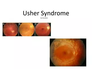
Retinosa Pigmentosa There a many varieties of RP, each with its own time of onset, degree of severity and set of symptoms.The overall uniting characteristic is the decay of photoreceptor cells, which are found in the retina and include two types – rods and cones. Whereas these cells die off naturally in all people, those with RP do not generate new rods and cones as would a healthy retina. The result is a slow decay of vision.
Symptoms of Retinosa Pigmentosa The optic nerve (arrow) looks very pale, the vessels (stars) are very thin and there is characteristic pigment. Pigmentation starts in periphery and gets closer towards the central vision. • Decreased night vision • Light sensitivity • Bad adaptation when moving from bright to dark • Visual field loss (tunnel vision/blind spots) www.nidcd.nih.gov/health/hearing/usher.asp