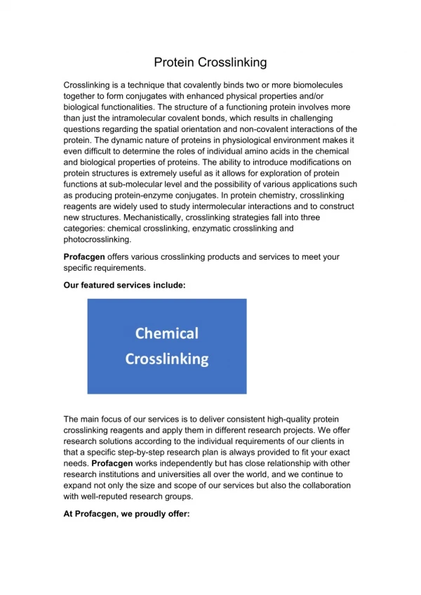
CROSSLINKING OF PROTEINS
40 likes | 212 Vues
CROSSLINKING OF PROTEINS. REFERENCES: Reithmeier , R.A.F. and Bragg, P.D. (1977). Biochim Biophys Acta . 466:245-256. Angus and Hancock. (1983). Bacteriol . 153:1042-51. . Kris-itd.unair.ac.id (for education purpose only). MATERIALS:

CROSSLINKING OF PROTEINS
E N D
Presentation Transcript
CROSSLINKING OF PROTEINS REFERENCES: Reithmeier, R.A.F. and Bragg, P.D. (1977). BiochimBiophysActa. 466:245-256. Angus and Hancock. (1983). Bacteriol. 153:1042-51. Kris-itd.unair.ac.id (for education purpose only)
MATERIALS: Outer membranes (or purified proteins) at a protein concentration of approx. 2 mg/ml (500 µg/ml). dithio-bis-succinimidylpropionate (DSP) stock solution; make up in DMSO at 15X the desired final concentraion (Final concentration will be approx. 2 mg/ml for outer membranes and 0.5 mg/ml for purified proteins) 0.2 M Triethanolamine buffer pH 8.5 1 M Tris-HCl pH 8.5 gel sample buffer without ß-mercaptoethanol (2-ME) (ie. 4% SDS/0.5 M Tris pH 6.8/20% gylcerol). heating block at 100 oC 11% ployacrylamide gel Second dimension; soaking buffer: 0.125 M Tris-HCl pH 6.8/10% 2-ME or 0.125 M Tris-HCl pH 6.8/10% dithiothreitol. 11% gel stacking gel with no wells ( poured to 3/16 from top of plate). We also have a special comb which incorporates two small wells to run molecular weight standards or a reference sample concurrently. 5 ml 0.8% agarose/gel * NOTE: - the number of gels you require for the second dimension should be made at the same time as the one for the first dimension.
METHOD: First dimension: outer membrane and purified protein preparations are prepared in 0.015 ml volumes of 0.2 M triethanolamine buffer pH 8.5, following the specifications of Reithmeier and Bragg (1977). Add 1 µl of the crosslinker, DSP, ( dissolved in dimethylsulphoxide) to make a 1/15 dilution to give the optimun final concentration. For the first experiment, a range of concentrations should be tried. React at room temperature for between 15 s and 2 min ( also determined by trial). A shorter reaction time would be preferable, to minimize fortuitous crosslinking of protein. Add 5 µl 1 M Tris-HCL pH 8.5 (an excess) to stop the crosslinking reaction. Dilute 1:1 into sample buffer containing SDS, Tris and glycerol without reducing agent. Heat samples at 100oC for ten minutes. Electrophoresis in the first dimension as for a normal protein gel. It is best to run parallel samples for second dimension electrophoresis on one side of the gel and those for direct staining to visualize the first dimension on the other side.
Second Dimension: Cut strips representing each sample well to be re-electrophored from the first dimension gel with a razor blade. We have special blades which are 5 in. long for this purpose. Make a very small notch to mark the bottom end of each cut out strip before soaking them. Soak the first dimension gel strips in 10% 2-mercaptoethanol (v/v) or 10mM dithioerythritol (w/v) in a plastic petri dish for 30 min at room temperature, with intermittent agitation. Lay the soaked strips carefully atop the second dimension gel, being sure to pre-moisten the glass of the gel plates with electrophoresis buffer so that the strips will slide between the plates. Using gloves and a thick spacer, nudge the strips into the gap between the plates so that the strip is in contact with the second gel at all points along its length. Mark which end is the top of the strip and which is the bottom on the plates. Seal the strips atop the second dimension SDS-polyacrylamide gel with 0.8% (w/v) agarose and electrophorese as normal. A small amount of the same sample (or tracking dye) may be added if a well was incorporated into the stacking gel. This will serve as a reference point to find the protein(s) you are interested in (and/or to follow the progress of the run).