Download
1 / 40
520 likes | 1.15k Vues
Pregnancy and Lactation. DR. ZAHOOR ALI DR. AMEL EASSAWI. Fertilization . Normal site of fertilization is ampulla of oviduct upper lateral third of oviduct Must occur within 24 hours after ovulation Viability of ovum – 24 hrs .

E N D
Pregnancy and Lactation DR. ZAHOOR ALI DR. AMEL EASSAWI
Fertilization • Normal site of fertilization is ampulla of oviduct upper lateral third of oviduct • Must occur within 24 hours after ovulation • Viability of ovum – 24 hrs. • Sperm usually survive about 48 hours but can survive up to 5 days in female reproductive tract • Sperm deposited in vagina travel through cervical canal, uterus, and to upper third of oviduct • Female reproductive tract aids in sperm migration • Contractions of myometrium • Upward contractions of oviduct smooth muscle
Fertilization • First sperm to reach ovum • Sperm binds with specific site on zona pellucida on ovum • The sperm undergo the “Acrosomalreaction” and secretes powerful enzymes that helps it to pierce through the corona radiata and zona pellucida of the ovum. • Fuses with plasma membrane of ovum • This fusion Triggers chemical change in ovum’s surrounding membrane that makes outer layer impermeable to entry of any more sperm • Head of fused sperm gradually pulled into ovum’s cytoplasm • Within hour, sperm and egg nuclei fuse • Fertilized ovum now called a zygote
IMPORTANT • Only sperm of same species can bind with zonapellucida ( glycoprotein) of ovum. In human it has ZP3 receptors.
Fertilization and Implantation • Fertilized ovum( zygote ) having 46 chromosomes divides mitotically • Within week zygote grows and differentiates into blastocyst capable of implantation • The blastocyst implants in the posterior wall of uterus on 6-7th day after fertilization. • The blastocyst has inner cell mass that becomes embryo and outer cellular layer, Trophoblast (fetal portion of placenta). • At site of implantation Endometrium transforms into Decidual layer.(maternal portion of placenta).
Early Stages of Development from Fertilization to Implantation
Fertilization and Implantation • Blastocyst implants in endometrial lining by means of enzymes released by trophoblasts • Trophoblastic layer in embryo is called Chorion
Formation of Placenta • After implantation Placenta develops • Placenta forms link between the fetus and mother. • Trophoblast(chorion) forms fetal part • Decidual layer of the endometrium forms the maternal part • This entire system of Decidual • ( mother) and chorionic (fetal) • Structures make placenta.
Formation of Placenta • We have finger like projections of chorionic tissue in the pool of maternal blood. • Embryo sends out capillaries in the chorionic projections to form placental villi • Each placental villus contains capillary surrounded by chorionic tissue • Chorionic tissue separates fetal blood from Maternal blood and works as barrier between the two.
Formation of Placenta • Placenta is fully operational by 5th week. • Throughout gestation blood flows between the placental villi and circulatory system of fetus by means of two umbilical arteries and one umbilical vein, which are wrapped in umbilical cord, a life line between fetus and placenta • Maternal blood is continuously replaced as fresh blood enters through uterine arterioles.
Functions of Placenta • Placenta performs the functions of Digestive system, Respiratory system and Kidney for the fetus. • Nutrition and O2 move to fetus from maternal blood across the placental barrier. • APPLIED Many drugs, chemical agents and micro organism can cross the placental barrier and harm the fetus (Aspirin, alcohol, cigarette smoking).There is History of babies born without limbs due to exposure to Thalidomide, a tranquilizer which was prescribed for pregnant women
Functions of Placenta • Organ of exchange between maternal and fetal blood • Acts as transient, complex endocrine organ that secretes essential pregnancy hormones 1. Human chorionic gonadotropin( HCG) 2. Estrogen 3. Progesterone 4. Human Chorionic Somatotrophin
1. Human chorionic gonadotropin(HCG) • Peptide hormone that prolongs the life span of corpus luteum • Appears in blood within 1 week and urine within 2 weeks of conception. • HCG -Reaches peak at 8-10 weeks then declines Actions of HCG : • Similar to LH. Maintains Corpus Luteum of pregnancy to secrete Progesterone. At 8-10 weeks Placenta is fully developed and takes over the secretion of Progesterone. • In male fetus-stimulate secretion of testosterone • Presence in urine forms the basis of pregnancy test.
2. Estrogen • 1st trimester – secreted from Corpus Luteum • After that Placenta secretes Estriol. • Why developing placenta doesn’t produce estrogen? • As Placenta lacks certain enzymes needed for synthesis of estriol • formation of Estriol requires the participation of Fetal Adrenal cortex together with Placenta. Actions During Pregnancy:- • Stimulates growth of myometrium--increasing uterine strength for parturition • Promotes development of ducts in mammary gland-prepare for lactation .
secretion of estrogen & progesterone from placenta The Feto-Placental Unit
3. Progesterone • 1st Trimester – secreted from Corpus Luteum: after that from Placenta. Actions During Pregnancy – • Inhibits uterine contractions“Hormone of pregnancy”. • Stimulates alveolar development in Mammary gland- prepares for lactation . • Promotes formation of mucus plug in cervical canal – prevention of uterine contamination.
4. Human Placental Lactogen(HPL) /Human Chorionic Somatotrophin • Protein hormone secreted by syncytiotrophoblast • Similar in structure to GH and Prolactin Actions – • Mobilizes nutrients from mother to fetus. • Helps prepare the mammary glands for lactation (similar to prolactin)
Physical Changes within Mother to Meet Demands of Pregnancy • Period of pregnancy -About 40 weeks(counted from 1st day of last menstrual period). • Uterine enlargement • Breasts enlarge and develop ability to produce milk • Volume of blood increases 30% • Weight gain(Increased 10-15 Kgs) • Increased Heart Rate & Cardiac Output – 30-40% • Respiratory activity increases by about 20% • Urinary output increases • Kidneys excrete additional wastes from fetus • Nutritional requirements increase • Mother needs calcium for calcification of fetal bones.
Parturition or Labor • Labor, delivery, birth • Requires • Dilation of cervical canal to accommodate passage of fetus from uterus through vagina and to the outside • Contraction of uterine myometrium that are sufficiently strong to expel fetus • Exact factors triggering increase in uterine contractility and initiating parturition not fully established
Factors That Triggers The Onset Of Labor • Role of high Estrogen levels: Rising estrogen level with gestation • Makes the uterus more excitable by • Increase the number of gap junctions between myometrial cells • Increases oxytocin receptors in myometrium • contributes to cervical softening by • Increase local prostaglandin production
Factors That Triggers The Onset Of Labor • Role of Oxytocin: Produced by hypothalamus, & released by posterior pituitary on neural stimulation • A powerful uterine muscle stimulant • Uterine responsiveness to oxytocin is 100 times greater at term than in nonpregnant women (because of the increase in gap junctions and increased concentration of myometrial oxytocin receptors) • labor is initiated when the myometrial responsiveness to oxytocin reaches a critical thresholdthat permits the onset of strong, coordinated contractions in response to ordinary levels of circulating oxytocin.
Factors That Triggers The Onset Of Labor • Role of Corticotropin Releasing Hormone(CRH): • secreted by the fetal portion of the placenta into both the maternal and fetal circulations • when a critical level of placental CRH is reached, parturition is triggered.
Parturition • Once contractions begin at labor onset, positive-feedback cycle progressively increases force of contraction • Pressure of fetus against cervix reflexly increases oxytocin secretion. • Which further pushes the fetus against the cervix • This cycle is reinforced as oxytocin stimulates prostaglandin secretion by decidua.
Stages of Labor • Cervical dilation ( first stage) • Longest stage, cervix dilates up to 10 cm • Lasts from several hours to as long as 24 hours in a first pregnancy • Delivery of baby(second stage) • Begins when cervical dilation is complete • Stretching of vagina causes reflex contraction of abdominal muscles • Usually lasts 30 to 90 minutes • Delivery of placenta( third stage) • Second series of uterine contractions separates placenta from myometrium • Shortest stage – usually completed within 15 to 30 minutes after baby is born
Stages of labor • IMPORTANT – If baby head is not down at the cervix but any other part eg feet , it is called Breech presentation which will not cause effective dilation of cervix, such cases require medical intervention eg Forceps delivery, caesarian section UTERINE INVOLUTION • After delivery, uterus shrinks to pregestational size (involution) in 4-6 week • During involution , remaining endometrium is expelled producing vaginal discharge called LOCHIA, for 3-6 weeks after delivery.
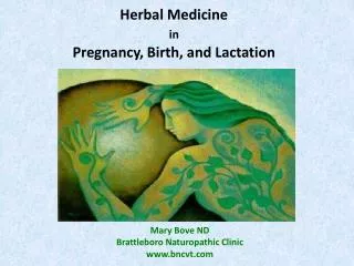
Lactation • Structure of mammary gland • Made up of lobules which contain alveoli/acinilined by epithelial cells. • Myoepithelial cells lie in between. • Ducts arise from each alveoli and converge to form lactiferous duct which open on the nipple.
Preparation of Mammary Gland for Lactation Hormones Acting on Mammary Gland • Estrogen : Ductal development. • Progesterone– Causes alveolar development. • Prolactin- Lactogenic hormone responsible for milk secretion. • Oxytocin – causes milk ejection ( let down). • Other hormones like cortisol, insulin , parathyroid hormone& growth hormone are also necessary for milk production.
Prevention of Lactation During Gestation • During pregnancy estrogen, progesterone and prolactin prepare alveoli for milk production. But actual milk flow takes place after parturition. • Why ?
Prevention of Lactation During Gestation Blocks PROLACTIN Stimulatory action High Estrogen & Progesterone during pregnancy Milk Secretion • At parturition sudden fall of estrogen and progesterone initiates lactation
Stimulation of Lactation by Suckling • Oxytocin is necessary to causes contraction of myoepithelial cells – milk ejection. Oxytocin secretion increased by suckling reflex. • Suckling also increases prolactin secretion which maintains milk production. • Prolactin ----- Promotes Milk Secretion • Oxytocin------ Milk Ejection
Factors Affecting Lactation • Adrenaline inhibits contraction of myopeithelial cells. Therefore emotional disturbances will inhibit milk ejection. • Other stimuli e.g. Crying of infant etc. can stimulate milk flow(condition reflex). • During nursing Menstrual cycle is absent in mother due to high levels of Prolactin which inhibit Pituitary Gonadotrophins - Locational Amenorrhea
References • Human Physiology, Lauralee Sherwood, seventh edition. • Text book Physiology by Guyton &Hall,11th edition. • Text book of Physiology by Linda s. Contanzo, third edition. • Physiology by Berne and Levy, sixth edition.