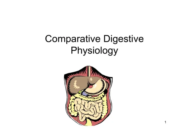
ALIMENTARY TRACT
ALIMENTARY TRACT. Histology A Text and Atlas; 6. th Edition, Michael H. Ross & Wojciech Pawlina (LWW) Chapter 17; Page: 568-627. OVERWIEV OF THE DIGESTIVE TRACT.

ALIMENTARY TRACT
E N D
Presentation Transcript
ALIMENTARY TRACT Histology A Text and Atlas; 6.th Edition, Michael H. Ross & Wojciech Pawlina (LWW) Chapter 17; Page: 568-627
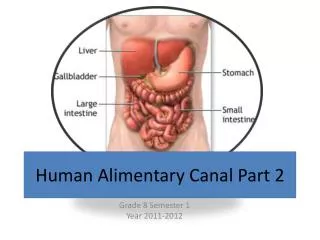
OVERWIEV OF THE DIGESTIVE TRACT • The portion of the alimentary canal that extends from theproximal part of the esophagus to the distal part of the analcanal is a hollow tube of varying diameter. • This tube has thesame basic structural organization throughout its length. • The Digestive Tract is composed basically of FOUR major parts or organs • A. EOSOPHAGUS • B. STOMACH • C. SMALL INTESTINE • D. LARGE INTESTINE • All parts separated one from another by muscular valves or sphincters.
OVERWIEV OF THE DIGESTIVE TRACT • All parts show FOUR layers or tunicae • THE MUCOSA, consisting of a lining epithelium, an underlyingconnective tissue called the lamina propria, and the muscularismucosae, composed of smooth muscle. • THE SUBMUCOSA, consisting of dense irregular connective tissue • THE MUSCULARIS EXTERNE, consisting in most parts of two layers of smooth muscle. • THE SEROSA or ADVENTITIA: • Serosa, a serous membrane consisting of a simple squamous epithelium, the mesothelium, and a small amount ofunderlying connective tissue. • An adventitia consistingonly of connective tissue is found where the wall of thetube is directly attached or fixed to adjoining structures(i.e., body wall and certain retroperitoneal organs).
LAYERS SPECIFICATION : MUCOSA • The structure of the alimentary canal varies considerably from region to region; • Generally, this variationoccurs within the mucosa. • The epithelium differsthroughout the alimentary canal and is adapted to the specificfunction of each part of the tube. • The mucosa has three principalfunctions: • Protection • Absorption • Secretion • The mucous membrane ( tunica mucosa) consist ofthe following layers: • a. SURFACE EPITHELIAL MEMBRANE : It is wet, lubricated by mucus and restin upon a basal lamina. • b. THE LAMINA PROPRIA : It is supporting layer. It is composed of LOOSE AREOLAR CONNECTIVE TISSUE. • c. THE MUSCULARIS MUCOSA : It is thin. Ussually arranged in two layers oriented as inner circular and outer longitudinal .
EPITHELIUM OF MUCOSA: BARRIER FUNCTION • The epithelial barrier separates the external luminal environmentof the tube from the tissues and organs of the body. • Thebarrier aids in protection of the individual from the entry ofantigens, pathogens, and other noxious substances. • In theesophagus, a stratified squamous epithelium provides protectionfrom physical abrasion by ingested food. • In the gastrointestinalportion of the alimentary tract, tight junctionsbetween the simple columnar epithelial cells of the mucosaserve as a selectively permeable barrier.
EPITHELIUM OF MUCOSA: ABSORPTION FUNCTION • The absorption of digested nutrients, water, and electrolytesis possible because of projections of the mucosa and submucosainto the lumen of the digestive tract. • These surface projectionsgreatly increase the surface area available forabsorption. • They consist ofthe following structural specializations: • Plicae circularesare circumferentially oriented submucosalfolds present along most of the length of the small intestine. • Villi are mucosal projections that cover the entire surface ofthe small intestine, the principal site of absorption of the products of digestion. • Microvilli are tightly packed, microscopic projections ofthe apical surface of intestinal absorptive cells. • In addition, the glycocalyxconsists of glycoproteins. It is essential for the final steps of digestion of proteins and sugars.
EPITHELIUM OF MUCOSA: SECRETION FUNCTION • Secretion is carried out largely by glands distributed throughoutthe length of the digestive tube. • The various secretoryproducts provide protection, buffering, assist in digestion and defence. • Mucus:protectivelubricationandbuffering • Enzymes, hydrochloric acid, peptide hormones,and water: assistin digestion, • Antibodiessecretion: Defence
GLANDS OF ALIMENTARY TRACT • The glands of the alimentary tract developfrom invaginations of the luminal epithelium and include • mucosal glands that extend into the lamina propria. • submucosalglands that either deliver their secretionsdirectly to the lumen of mucosal glands or via ducts thatpass through the mucosa to the luminal surface. • extramural glands that lie outside the digestive tract anddeliver their secretions via ducts that pass through the wallof the intestine to enter the lumen. • The liver and the pancreasare extramural digestive glands. • They deliver their secretions into the duodenum.
LAMINA PROPRIA • The lamina propria contains • Gland, • Vessels (transport absorbed substances), • Component of the immune system (Lymphatic Tissues). • Aggregetion of mucussecretingglands (lubricatethe epithelial surfaceto protect the mucosa frommechanical and chemical injury) • Typically, the blood capillariesare of the fenestrated type and collect most of the absorbedmetabolites. • In the small intestine, lymphatic capillaries are numerousand receive some absorbed lipids and proteins. • The lymphatic tissues in the lamina propria function asan integrated immunologic barrier that protects againstpathogens and other antigenic substances. • The lymphatic tissue is represented by • diffuse lymphatic tissue • lymphatic nodules with well-developed germinal centers. • eosinophils, macrophages, and sometimes neutrophils. • The diffuse lymphatic tissue and the lymphatic nodules arereferred to as gut-associated lymphatic tissue (GALT). • The ileum, extensive aggregates ofnodules, called Peyer’s patches, • Aggregatedlymphatic nodules are also present in the appendix.
MUSCULARIS MUCOSA • The muscularis mucosae, the deepest portion of themucosa, consists of smooth muscle cells • This layer arrangedin an innercircular and outer longitudinal layer. • Function: Contraction of this muscleproduces movement of the mucosa, forming ridges andvalleys that facilitate absorption and secretion.
LAYERS SPECIFICATION: THE SUBMUCOSA • The submucosa consists of a dense, irregular connectivetissue • This layer contains: • Blood andlymphatic vessels, • A nerve plexus, • Glands. • The extensive nerve network in the submucosa contains visceralsensory fibers mainly of sympathetic origin, parasympathetic(terminal) ganglia, and preganglionic and postganglionicparasympathetic nerve fibers. • The nerve cell bodies of parasympatheticganglia and their postganglionic nerve fibers representthe enteric nervous system. • In the submucosa, the network of unmyelinatednervefibers and ganglion cells constitute the submucosalplexus(also called Meissner’s plexus). • As noted, glands occur occasionally in the submucosaincertain locations. • For example, they are present in the esophagusand the initial portion of the duodenum.
LAYERS SPECIFICATION: MUSCULARIS EXTERNA • In most parts of the digestive tract, the muscularisexternaconsists of two concentric and relatively thick layers ofsmooth muscle. • The cells in the inner layer form a tight spiral,described as a circularly oriented layer; • Those in theouter layer form a loose spiral, described as a longitudinallyoriented layer. • Located between the two muscle layers is athin connective tissue layer. • Within this connective tissue liesthe myenteric plexus (also called Auerbach’s plexus),containing nerve cell bodies (ganglion cells) of postganglionicparasympathetic neurons and neurons of the enteric nervoussystem. • Contractions of the muscularisexterna mix and propelthe contents of the digestive tract. • Contraction of the inner circular layer of the muscularisexternacompresses and mixes the contents by constricting thelumen; • Contractionof the outer, longitudinal layer propelsthe contents by shortening the tube. • The slow, rhythmiccontraction of these muscle layers under the control of the entericnervous system produces peristalsis (i.e., waves of contraction). • Peristalsis is marked by constriction and shortening ofthe tube, which moves the contents through the intestinal tract.
LAYERS SPECIFICATION: MUSCULARIS EXTERNA • A few sites along the digestive tube exhibit variations in themuscularisexterna. • In the wall of the proximalportion of the esophagus (pharyngoesophageal sphincter) andaround the anal canal (external anal sphincter), striatedmuscleforms part of the muscularisexterna. • In the stomach, athird, obliquely oriented layer of smooth muscle is presentdeep into the circular layer. • Finally, in the large intestine, partof the longitudinal smooth muscle layer is thickened toform three distinct, equally spaced longitudinal bands calledteniaecoli.
LAYERS SPECIFICATION: MUSCULARIS EXTERNA • The circular musclelayer is thickened to form sphincters or valvesatseveral points along the digestive tract. • From theoropharynx distally, these structures include the following: • Pharyngoesophageal(superior - upper) esophageal sphincter: • Located: The lowestpart of the cricopharyngeusmuscle • Function: Preventsthe entry of air into the esophagus. • Inferior (lower) esophageal sphincter: • Located: At the lower end of the esophagus, • Function: Prevents reflux of gastric contents into theesophagus • Pyloric sphincter(gastroduodenal sphincter): • Located:At the junction of the pylorusof the stomach and duodenum • Function: Controlsthe release of chyme, the partially digestedcontents of the stomach, into the duodenum. • Ileocecal valve: • Located:At the junction of the small andlarge intestines, • Function: Prevents reflux of the contents of thecolon with its high bacterial count into the distal ileum. • Internal anal sphincter: • Located: This, the most distally locatedsphincter, surrounds the anal canal • Function: Prevents passage ofthe feces into the anal canal from the undistended rectum.
LAYERS SPECIFICATION: SEROSA or ADVENTITIA • SEROSA: • The serosa is a serous membrane consisting of a layer ofsimple squamous epithelium, called the mesothelium, and asmall amount of underlying connective tissue. • Theserosa is the most superficial layer of those parts of the digestivetract that are suspended in the peritoneal cavity. • The serosa is continuous with both the mesentery and thelining of the abdominal cavity. • Large blood and lymphatic vessels and nerve trunkstravel through the serosa. • Large amounts of adiposetissue can develop in the connective tissue of the serosa (and in the mesentery). • ADVENTITIA: • The thoracic part of the esophagus and portions of structuresin the abdominal and pelvic cavities are fixed to thecavity wall such as the duodenum, ascending and descendingcolon,rectum, and anal canal. • These structures are attached to theabdominal and pelvic wall by connective tissue.
ESOPHAGUS: FUNCTION AND ANATOMY • The esophagus is a fixed muscular tube. • Function: The esophagus deliversfoodand liquid from the pharynx to the stomach. • Anatomy: • The overall length of the esophagusis about 25 cm. • The neck andmediastinum region of esophagus is attached to adjacent structures by connective tissue (Adventitia). • The abdominal cavity region of esophagus is free (approximately 1 to 2cm) (Serosa). • On cross section, the lumen normally collapsed state • Esophagal lumen has a branched appearance because oflongitudinal folds. • When a bolus of food passes through theesophagus, the lumen expands without mucosal injury.
HISTOLOGY OF ESOPHAGUS: MUCOSA • The mucosa that lines the length of the esophagus has anonkeratinized stratified squamous. • In humans,the surface cells may exhibit some keratohyalin granules, • Keratinization does not normally occur. • The underlyinglamina propria is similar to the lamina propria throughoutthe alimentary tract: • Diffuse lymphatic tissue (DLT)and lymphatic nodules, • The esophageal mucous glands. • Themuscularis mucosa,is composed of longitudinally organized smooth musclethat begins near the level of the cricoid cartilage. • Functions:Aid in swallowing.
HISTOLOGY OF ESOPHAGUS: SUBMUCOSA • The submucosa consists of dense irregular connectivetissue • This layer contains the larger blood and lymphatic vessels,nerve fibers, and ganglion cells. • The nerve fibers and ganglioncells make up the submucosal plexus (Meissner’s plexus). • Glands are also present. • Glands and DLTare more prevalent inthe upper and lowerparts of the esophagus • In addition, diffuselymphatic tissue(DLT) and lymphatic nodules are present.
HISTOLOGY OF ESOPHAGUS: MUSCULARIS EXTERNA • The muscularis externaconsists of two muscle layers: • Aninner circular layer • Anouter longitudinal layer. • It differs from the muscularis externa found in therest of the digestive tract • The upper one third is striatedmuscle, a continuation of the muscle of the pharynx. • The muscularis externa of the middle thirdis mixed (Striatedand Smoothmuscle) • The muscularis externa of the distal third consists only ofsmooth muscle. • A nerveplexus, the myenteric plexus (Auerbach’s plexus), is presentbetween the outer and inner muscle layers. • Nerve plexus innervates the muscularis externaand produces peristaltic activity.
GLANDS OF THE ESOPHAGUS • Function: Mucosal and submucosal glands of the esophagus secretemucus to lubricate and protect the luminal wall. • Two types galnds are present in the wall of the esophagus. • Esophageal glands proper: • Liein the submucosa. • More concentrated in the upper half. • Small, compound, tubuloalveolarglands. • Esophageal cardiac glands (name similarity to the cardiac glands of the stomach) • Foundin the lamina propria of the mucosa. • Present inthe terminal part of the esophagus.
INNERVATION OF ESOPHAGUS • The muscle of the esophageal wall is innervated bybothautonomic and somatic nervous systems. • The striated musculature in the upper part of the esophagusis innervated by somatic motor neurons of the vagus nerve,(from the nucleus ambiguus). • The smooth muscleof the lower part of the esophagus is innervated by visceralmotor neurons of the vagus (from the dorsal motor nucleus).
CLINICAL CORRELATION: GASTROESOPHAGAL REFLUX DISEASE • Inferior (lower) esophageal sphincter prevents reflux of gastric contents into theesophagus. • Esophagal glands near the stomach tend to protectthe esophagus from regurgitated gastric contents. • Abnormal relaxation of inferior esophagealsphincter allowsacidic content of the stomach to return (reflux) into theesophagus. • Esophagal glands are not fully effective, • If not treated, this conditions may progress intogastroesophageal reflux disease (GERD), • GERD is characterizedby inflammation of the esophageal mucosa (refluxesophagitis), strictures, and difficulty in swallowing (dysphagia) with accompanying chest pain (heartburn) • Excessivereflux results in pyrosis, • The muscle of the esophageal wall is innervated by bothautonomicand somatic nervous systems.
STOMACH: FUNCTION AND ANATOMY • Localization: The stomach is an expanded part of the digestive tubethat liesbeneath the diaphragm. • Function: It receives the bolus of macerated foodfrom the esophagus. • Mixing and partial digestion of the food inthe stomach by its gastric secretions • Stomach produce a pulpy fluid mixcalled chyme. • Anatomy: Gross anatomists subdivide the stomach into four regions. • The cardia: surrounds the esophageal orifice; • The fundus: liesabove the level of a horizontal line drawn through theesophageal (cardiac) orifice; • The body lies below this line; • The pyloric part (funnel-shaped)is the region that leads into thepylorus, the distal, narrow sphincteric region between thestomach and duodenum.
HISTOLOGIC REGION OF THE STOMACH • The stomach is subdivided into only three regionsbyhistologist. • These subdivisionsare based on the types of glands. • The histologic regions are as follows: • Cardiac region (cardia), the part near the esophageal orifice, • Pyloric region (pylorus), the part proximal to the pyloricsphincter, • Fundic region (fundus), the largest part is situated between the cardia and pylorus.
General Specification of the Gastric Mucosa • The stomach has the same general structural plan throughout. • The stomach consisting of a mucosa, submucosa, muscularis externa, and serosa. • Rugae: Examination of the inner surface of the emptystomach reveals a number of longitudinal folds or ridgescalled rugae. • Localization: Prominent in the narrower regions ofthe stomach. • Functions: Serve to accommodate expansion and filling of the stomach.
General Specification of the Gastric Mucosa • Mamillated Areas: Smaller regions of the mucosa are formed by grooves orshallow furrows that divide the stomach surface intobulging irregular areas called mamillated areas. • Functions: Provide a slightly increased surface area for secretion. • Gastric pits (foveolae) At higher magnification, numerous openings can be observedin the mucosal surface called foveolae. • Functions: The gastric glands openinto the bottom of the gastric pits.
SURFACE MUCOUS CELL • Surface mucous cells is found in the inner surface of the stomach and the gastric pits. • The epithelium is simple columnar and secrete the visible mucus. • Each cell has a large, apical cup of mucinogen granules. • It typically appears empty in routine H&E sections. • The mucinogen granules stain intensely with toluidine blue and with the periodic acid–Schiff (PAS) procedure.
Protection of the Gastric Mucosa • Protection Factors: • Gasttric mucus, • High bicarbonate and potassium concentration, • Prostaglandins (PGE2) • Gastric mucus protection mechanism: • Thisforms a thick, viscous, gel-like coat. • This mucusadheres tothe epithelial surface, • Thus, it protects against abrasionfrom rougher components of the chyme. • High bicarbonate and potassium concentration protection mechanism: • The bicarbonate makes the mucus alkaline • The bicarbonate is not rapidly mixing from the contents of the gastric lumen because it is containment within the mucous coat. • Prostaglandins (PGE2) protection mechanism: • They stimulate secretion of bicarbonates • Theyincrease thickness of the mucus layer with accompanied vasodilatation in the lamina propria. • Vasadilatation improves supply of nutrients • PGE2 effect is provided optimizing conditions for tissue repair
CLLINICAL CORRELATION: Aspirin and NSAIDs • The lining of the stomach does not function in an absorptivecapacity. • However, some water, salts, and lipidsolubledrugs may be absorbed. • Alchol andcertain drugs such as aspirin ornonsteroidalanti-inflammatorydrugs (NSAIDs) enter the lamina propria by damagingthe surface epithelium. • Even small doses of aspirinsuppressthe production of protective prostaglandins bythe gastric mucosa. • In addition, aspirin’s direct contactwith the wall of the stomach interferes with the hydrophobicproperties of the gastric mucosa.
Fundic Glands of the Gastric Mucosa • Thefundic glands (gastric glands)produce the gastric juice of the stomach. • Thefundic glands are simple, branched, tubular glands. • The gastric glands extendfrom the bottom of the gastric pits to the muscularismucosae. • Typically, several glands open into a singlegastric pit. • The base of the gland usually divides into two and sometimesthreebranches. • Gastric glands is divided three segments (order from surface to base): • Isthmic segment: Short segment (site of stem cell location), • Neck Segment: Narrow, relatively long, • Fundic (Base) Segment: Shorter, wider and slightly coiled.
SECRETION of the FUNDIC GLANDS • The cells of the gastric glands produce gastricjuice (about 2 L/day). • Gastric juice contains four major components, water and electrolytes: • Hydrochloric acid (HCl), • Pepsin, • Mucus, • Intrinsic factor. • Hormones and hormonelike factors
SECRETION of the FUNDIC GLANDS • Hydrochloric acid (HCl): • It is produced by parietal cells • Provide the gastric juice a low Ph(1.0 to 2.0). • Initiatesdigestion of dietary protein. • Converts inactive pepsinogen intothe active enzyme pepsin. • Pepsin, a potent proteolytic enzyme. • It is produced by the chief cells • Pepsin hydrolyzes proteins into small peptidesby splitting interior peptide bonds. • Mucus: • Several types of mucus-producing cellssecrete an acid-protective coat • Physiologic gastric mucosa barrier: The mucus andbicarbonates trapped within the mucous layer maintain aneutral pH. • Physical barrier of mucosa: Mucus serves as abetween the cells of the gastric mucosa andthe ingested. • Intrinsic factor (a glycoprotein) • It is secreted by parietal cells • Intrinsic factor binds to vitamin B12. • Vitamin B12 It is essential for its absorption,which occurs in the distal part of the ileum. • Hormones (Gastrin)andhormonelikesecretions are produced by enteroendocrinecells. • This products secreted into the lamina propria, • Then, they enter the circulation or act locally on other gastric epithelial cells.
Cells of the Fundic Glands • Fundic glands are composed of four functionally different cell types. • Each cell has a distinctive appearance. • Undifferenriated cells give rise to all fundic gland cells. • Various cells constitute the gland: • Mucous neck cells • Chief cells • Parietal cells, also called oxyntic cells • Enteroendocrine cells • Undifferentiated adult stem cells
MUCOUS NECK CELLS • LOCALIZATION: The mucous neck cells are located inthe neck region of the fundic gland. • Parietal cells are usuallyinterspersed between groups of these cells. • STRUCTURE: The mucous neckcell hasshorterheight and spherical prominent nucleus. • Release of mucinogen granules is inducedby vagal stimulation; • Thus, secretion from these cellsdoes notoccur in the resting stomach.
CHIEF CELLS • LOCALIZATION: Chief cells are located in the deeper part of the fundic glands. • FUNCTION: Chief cells secretepepsinogen and a weak lipase • On contact with the acid gastricjuice, pepsinogen is converted to pepsin, a proteolytic enzyme. • STRUCTURE: Chief cells are typical protein-secreting cells. • The abundant rER in the basal cytoplasmgives this part of the cell a basophilic appearance, • The apical cytoplasm is eosinophilic owing to the presence of thesecretory vesicles (zymogen granules).
PARIETAL (OXYNTIC) CELLS • LOCALIZATION: Parietal cells are found in upper and middle portions of the neck. • FUNCTION: Parietal cells secrete HCland intrinsic factor. • STRUCTURE: They are large cells, triangular shape, sometimes binucleate, and spherical nucleus. • The cytoplasm stains with eosin and other acidic dyes. • Pariatal cells contain numerous mitochondria because of energy necessary for acid secretion. • On TEM examination: • Extensive intracellular canalicular system • Numerous microvilli project from the surface of the canaliculi, • An complicate tubulovesicular membrane system is present in the cytoplasm adjacent to the canaliculi. • The membranes of the tubulovesicular system serve as a reservoir of plasma membrane containing active proton pumps. • This membrane material (microvilli, intracellular canaliculi and tubulovesicular system) increase surface area and the number of proton pumps.
HCl Production • Parietal cells have three different types of membrane receptors • This receptors activate HCl secretion: • Gastrin receptors, • Histamine H2 receptors, • Acetylcoline M3 receptor. • The gastrin receptor is activated by gastrin, • Then, This activation stimulates parietalcell. • After stimulation, severalsteps occur in the production of HCl: • Production of H ions: • The parietal cell cytoplasm has the carbonic anhydrase enzyme. • This enzyme hydrolyzescarbonic acid (H2CO3) to H and HCO3. • Carbon dioxide (CO2) is requiredfor synthesis of carbonic acid, • CO2 diffusesacross the basement membrane into the cell from theblood capillaries. • Transport of H ions: • H ions passes from the cytoplasmto the lumen of the canaliculusby the H/K-ATPase proton pump. • Simultaneously, K from thecanaliculus is transported into the cell cytoplasm in exchange for the H ions. • Transport of K and Cl ions: • The parietal cellK and Cl channels (uniporters) is activated. • Formation of HCl from • TheH and Clthat were transportedinto the lumen of the canaliculus. • Then, HCl is produced in the lumen.
Intrinsic Factor • In humans, intrinsic factor is secreted by the parietal cells. • Its secretion is stimulatedby the same receptors that stimulate gastric acid secretion. • Intrinsic factor is a 44-kilodalton glycoprotein. • IF make complexe with vitamin B12 in the stomach and duodenum, • Vitamin B12 is absorbed from the ileum.
ENTEROENDOCRINE CELLS • Older names of the enteroendocrine cells are enterochromaffin cells, argentaffin cells, and argyrophil cells. • LOCALIZATION: • Enteroendocrine cells are found at every level of the fundic gland. • They are more prevalent in the base. • FUNCTION: • Enteroendocrine cells secrete their products into either the lamina propria or underlying blood vessels. • Two types of enteroendocrine cells can be distinguished throughout the gastrointestinal tract. • Enteroendocrine“closed” cells: Most of them represent small cells that rest on the basal lamina and do not always reach the lumen • Closed cells, is regulated by luminal content indirectly through neural and paracrine mechanisms. • Enteroendocrine“open” cells: Someof them have a thin cytoplasmic extension bearing microvilli that are exposed to the gland lumen • Open cells serve as primary chemoreceptors.
ENTEROENDOCRINE CELLS • STRUCTURE: • Electron micrographs reveal small membrane-boundsecretory vesicles throughout the cytoplasm. • With the aid of the TEM, at least 17 different types ofenteroendocrine cells have been described on the basis of size,shape, and density of their secretory vesicles. • The more than 20peptide and polypeptide hormones and hormone-like regulatingagentsare currently identified and characterizedby immunochemical staining.
Cardiac Glands of the Gastric Mucosa • LOCALIZATION: • Cardiac glands are found in the cardia region of stomach. • FUNCTION: • The stomach cardiac gland and the esophageal cardiac glandshelps protect the esophageal epithelium against gastric reflux. • STRUCTURE: • Cardiac glands are composed mainly of mucus-secreting cells, with occasional interspersed enteroendocrine cells. • The glands are tubular, convoluted, and occasionally branched. • The glands drain into shallow gastric pits • A short duct segment containing columnar cells. • The mucus-secreting cells are similar in appearance to the cells of the esophageal cardiac glands. • They have a flattened basal nucleus, and the apical cytoplasm is typically filled with mucin granules.
Pyloric Glands of the Gastric Mucosa • LOCALIZATION: • Pyloric glands are located in the pyloric antrum (the partof the stomach between the fundus and the pylorus). • FUNCTION: • Pyloric gland cells are help protect the pyloric mucosa. • STRUCTURE: • Pyloric glands arebranched, coiled, tubular. • The glands empty into deep gastric pits. • The lumen is relatively wide • Thesecretory cells are similarin appearance to the surface mucous cells, suggesting a relativelyviscous secretion. • Enteroendocrine cells are found interspersedwithin the gland epithelium.
EPITHELIAL CELL RENEWALL IN THE STOMACH • The isthmus of the fundic gland contains areservoir of tissue stem cells . • Surface mucous cells are renewed approximately every 3 to 5 days. • Most of the newly producedcells at this site become surface mucous cells. • They migrate upwardalong the wall of the pit to the luminal surface of the stomach • They are ultimately shed into the stomach lumen. • The cells of the fundic glands have a relatively long lifespan. • Other cells from the isthmus migratedown into the gastricglands • This cells give rise to the parietal cells, chief cells, mucousgland cells, and enteroendocrine cells • The mucous neck cellhas a much shorter lifespan, approximately 6 days. • The chief andenteroendocrinecells are estimated to livefor about 60 to 90days. • Theparietal cells have the longest lifespan, approximately 150 to200 days.
LAMINA PROPRIA • The lamina propria of the stomach is relatively scant and restrictedto the gastric pits andglands. • The stroma is composed largely of reticular fibers withassociated fibroblasts,smooth muscle and immune sytem cells. • Neutrophils may be prominent when inflammation occurs. • Lymphatic nodules are also present.