Download
1 / 16
170 likes | 366 Vues
MOLECULAR GENETICS OF CANCER-G PATHOLOGY {S1}. BY RANJEET RAMAN.

E N D
MOLECULAR GENETICS OF CANCER-G PATHOLOGY {S1} BY RANJEET RAMAN
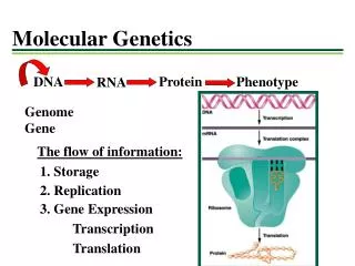
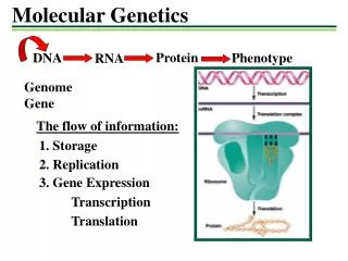
Evolution of unicellular to multicellular life Mutational load of a complex organism is hard to manage. Must separate germline early in embryology to protect it from excessive damage. Average human gene is mutated 1010 times in a lifetime! Nearly all are somatic mutations. • Innate resistance to tumorigenesis because single mutations are inadequate to cause cancer. Cancer has a multistep origin.
Neoplasia is a micro evolutionary process Clonal Selection Theory Numerous (5-7) rate-limiting mutations are required to cause cancer. Clonal Selection Theory says that tumorigenesis occurs as the serial expansion of successive growth-advantaged clones. One mutation is never sufficient because down regulation mechanisms always limit the effect.
Genetic heterogeneity exists even among the clone population, because of chromosomal instability and tendencies toward mutation. Subclones continue to mutate and compete in a sequential selection to dominate the neoplasm. • Subclones with the most extensive genetic change should have the worst prognosis. • Changes in stem cells and differentiation are required for neoplasia.
Pediatric tumors occur during the developmental window and are therefore quite distinct from tumors that develop in adults. They probably have far fewer mutations.
Types of mutations • 1. Amplification – leads to overproduction of protein. • 2. Rearrangement/translocation – fuses genes from different chromosomes, creating a novel protein or bringing an oncogene close to a strong promoter. • 3. Large deletion – total inactivation of a suppressor gene or loss of heterozygosity. This is bad. It is often the “second hit” that really screws the cell.
4.Viral insertion – allows continuous expression of viral oncogenes. • 5.Telomere shortening – may be a cause of genetic instability. In malignancy the telomerase reactivates.
There are three categories of mutated cancer genes: • 1. Dominant oncogenes (function activated by mutation. Normally transduce GF signals and oppose apoptosis) -Growth factors, which aren’t very oncogenic because neighbor cells benefit too.
Growth factor receptors. Example is EGFR. Over expression is very oncogenic. • Signal transducers, such as GTP-binding proteins (RAS) and tyrosin kinases (ABL). • Nuclear oncoproteins • Antagonists of apoptosis (Bcl2) • Antagonists of tumor suppression (MDM2, which antagonizes p53, a tumor- suppresor)
2. Tumor suppressor genes (function inactivated by mutation, providing immediate selective advantage) -cell/ECM interactions (e-cadherin) cell division cycle regulation -apoptosis (p53, BAX) 3. DNA-maintenance genes (function inactivated, providing selective advantage only if other oncogenic mutations occur) -DNA repair (xeroderma pigmentosum genes) -chromosome stability (Fanconi genes)
Inherited syndromes due to germline mutations, Mutations in germ line dominant oncogenes are very rare, because not well tolerated by embryo. • Mutations in germline tumor suppressor genes is a common cause of congenital cancer. Classic theory of cancer inheritance is the Nicholls Two-Hit Hypothesis, which states that inheriting one bad copy increases the rate of occurrence of neoplasm.
Mutations in DNA maintenance genes are true examples of highly mutating neoplasms. Chromosomal instability is very common. Best examples are xeroderma pigmentosum (inadequate repair of UV damage) and Hereditary Nonpolyposis Colorectal Carcinoma.
Rational Therapy • New drug screening strategies for specific biochemical properties New therapeutic strategies (augmenting deficient genes such as p53, replacing defected genes with good copies, inactivating bad genes, re-expressing silenced genes, activating immune system to kill tumors)
Molecular epidemiology and carcinogens • Dietary carcinogens • Infectious causes of cancer (indirect mechanisms of inflammation and mitogenesis such as with H.pylori and gastric cancer, or direct mechanisms such as HPV causing cervical cancer).
Defining a neoplasm • Neoplasms are clonal populations with autonomous growth and somatic mutations of growth-regulating genes. Neoplastic cells grow under normally limiting conditions. • Keloids, scars, and granulation tissue are NOT neoplasms. They are not clonal. Atherosclerotic plaques are clonal but not mutated, so they also are not neoplasms. • Most neoplasms are benign, and they are not always masses.
Neoplasms are not just proliferative in cell number (an example of this would be psoriasis). Rather, mutations cause exponential rises in the number of stem cells. This is cancer. • In colorectal cancer, the accumulation of many mutations is important, but their order isn’t. • In pancreatic cancer, a proper sequence of mutations occurs to derange cell cycle controls