Download
1 / 42
480 likes | 1.68k Vues
Safe Laparoscopic Access: technologies and techniques. 洪煥程 醫師 台北榮總 婦產部 96-1-2. Laparoscopic Abdominal Entry. Laparoscope-related complications were reported with increasing frequency during growing phase of laparoscopic surgery.

E N D
Safe Laparoscopic Access: technologies andtechniques 洪煥程 醫師 台北榮總 婦產部 96-1-2
Laparoscopic Abdominal Entry • Laparoscope-related complications were reported with increasing frequency during growing phase of laparoscopic surgery. • Entering the abdomen is the most dangerous part of the laparoscopic procedures.
Laparoscopic complications • Complications: • general-anesthesia-related • injuries to the vascular system, bladder, and bowel. • These injuries • indirect injury due to the use of monopolar current • now more commonly ascribed as direct trauma caused by the insertion of the insufflation needle or the primary or secondary trocars • Injuries occur more frequently as a result of the placement of the insufflating needle or primary trocar placement. Levy BS 1985; Reich H 1995
Laparoscopic complications • Most complications during laparoscopy occur during the surgeon’s first 100 cases. Soderstrom RM et al. Operative Laparoscopy: The Master’s Technique. 1993
Laparoscopic Abdominal Entry Approaches to initial cannula • Closed insertion without preinsufflation • Closed insertion with preinsufflation • Open laparoscopy
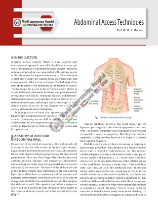
Anatomic landmarks • Umbilicus: at the level of L3 and L4 • Abdominal aorta: bifurcation L4 and L5
Umbilical area Lower edge Adequate size Complete horizontal position Placement of the Veress needle
Placement of the Veress needle • The angle insertion : 45 degree from the surface of skin, more vertical orientation in obese women • Aim toward uterus • Aim away from pelvic vessels (vertical; sagital) • Aim at right angle to the skin
Placement of the Veress needle • Palpating the aorta and sacral promontory • Grasp the base of umbilicus : keep Veress needle from the abdominal structure
Placement of the Veress needle • Using towel clips to elevate the abdominal wall
Tests of peritoneal insertion • Drop test (Epidural space test): - a drop of water --- suctioned into peritoneal cavity • Syringe barrel flow test: • Manometer test (free flow of gas): - elevation of abdominal wall, falling insufflation pressure ( < 5-6 mmHg at 1.0L/min flow ) • Loss of liver dullness early in insufflation
Tests of peritoneal insertion • Aspiration-instillation-aspiration test (Syringe aspiration test): - Aspiration : blood, bile, bowel content, urine - Instillation : 10cc N/S--- if any resistant - Re-aspiration : no fluid aspirated • Waggling test (Lateral needle swing test): (potentially dangerous) - base on tactile confirmation of a free- moving tip - pivot point at the tip --- adhesion or pre- peritoneal - pivot point at the fascia --- intraperitoneal
Recommended amount of gas for initial insufflation with a Veress needle
Primary trocar insertion • Patient in a horizontal position • Skin incision should be large enough to prevent any resistance • A stretched middle finger can prevent over-insertion (palming technique) • Z-technique ? (prevention of incision hernia)
Direct trocar insertionwithout preinsufflation • First described by Dingefelder (1978)
High risk conditions for abdominal entry • Strong abdominal musculature (sportswomen) • Body weight is obese or very thin • Large pelvic mass • Pregnancy • Previous abdominal and pelvic operations
The Incidence of Adhesions After Prior Laparotomy ? (N=360) Adhesion bowel omentum Pfannenstiel : 13% 87% Midline below the umbilicus : 17% 83% Midline above the umbilicus : 40% 60% Subjects with all types of incision : Gynecologic (42 %) > Obstetric (22%) Brill AL: Obs Gyn 1995, 85:269-72
Management of abdominal wall access with probable intraperitoneal adhesion • Exploratory syringe aspiration test • Direct Veress needle with optic catheter • Open laparoscopy • Alternative access
Exploratory syringe test(Syringe aspiration test) • To ascertain : no bowel/vessels adherent under the umbilicus before trocar insertion • After pneumoperitoneum created by Veress needle, a 20-gauge needle inserted under negative pressure at the four cardinal points of a 20-mm circle around the umbilicus --- alternate sites trocar if blood or bowel content is aspirated
Direct Veress needle insertion with optic catheter ( needle scopy) • 1.2mm, 1.75mm, 1.98 mm • Visual control via optics inserted into needle
Open laparoscopy • 1971, Hasson • A horizontal/vertical incision about 2 cm • Enter peritoneal cavity by incision of abdominal wall step by step • Apply Hasson trocar-cannula and purse-string suture at fascial level
Alternative access sites • inter/infracostal area : - the 9th intercostal space in the middle of the mid-clavicular & the ant axillary line • Lt costal margin : - 3-4 cm below the left costal margin in the mid-clavicular line - Reverse Trendelenburg position - First to rule out splenomegaly or insufflated stomach ( on NG )
Alternative needle insertion sites • Lt periumbilical area : - midclavicular line • Transvaginal insertion : - via post cervix fornix, trendelenburg position • Transabdominal insertion : - Pushed uterus up against the abdominal, then inserted through abdomen and into fundus of the uterus
Storz TERNAMIAN EndoTIP ( Endoscopic Threaded Imaging Port ) • Re-usable trocar • After umbilical incision and Veress insufflation, a 0° laparoscope is mounted in the cannula. The tip of the cannula is inserted into a tiny fascial incision and rotated clockwise • All the abdominal wall layers are well visualized
is used to safely create a Pneumoperitoneum in the scarred abdomen fascial incision should be 1 to 1.5 cm in size A long suture is placed on each fascial edges finger dissection a tunnel or an opening into the intraabdominal cavity is gently created The foamgrip anchoring device is set and secured with the previously placed suture Blunt Trocar
A 1 cm skin incision A telescope is inserted into the trocar and the path of entry of the trocar into intra-abdominal cavity is visualized These planes are cut slowly with the blade of the trocar (at the tip of the instrument) Pneumoperitoneum must be created or abdominal wall elevation must be performed prior to the insertion VISIPORT™
an integrated system combining a Nylon stretchable sheath over a Disposable Veress needle Once inserted, the sheath is dilated by inserting the trocar (with a dilator in place) no cutting entry blade decreasing trocar site bleed and the potential for an intra-abdominal injury creates a smaller fascial defect which does not need to be closed up to 12mm VERSASTEP SYSTEM™
Secondary trocar insertions • Off the midline, to the left, above the pubic hairline • Transillumination and under endoscopic direct vision helps to identify the vessels. and minimizing the risk of injury • Deep Inferior epigastric <-- ext. iliac • Superficial epigastric <-- femoral • Lateral to the rectus abdominis muscle • Lateral to the umbilical ligaments • Lateral to the deep epigastric vessels • Aiming toward the uterus (cul-de-sac) and away from the iliac vessels • Keeping the forefingers extended on the sleeve
Conclusion - 1 • The incidence and spectrum of access-related complications is greater than previously perceived. • Newer devices and modifications in technique may reduce the incidence of such adverse events.
Conclusion - 2 • Put patient in a proper position • Understand anatomical relation • Follow the abdominal entry principles • Be aware of high-risk conditions • Use proper instruments and alternative strategies
Q 1 • The angle insertion of Veress needle ? degree from the surface of skin ( more obese) 1. 30 degree 2. 45 degree 3. 90 degree 4. 0 degree
Q 2 • What vessel injury related to peritoneal hematoma during lat. trocarinsertion ? 1. Superficial circumflex iliac a. 2. Superficial epigastric a. 3. Deep circumflex iliac a. 4. Deep inferior epigastric a.