Download
1 / 19
190 likes | 490 Vues
E2 – Perception of stimuli. Human eye. Human ear. Human sensory receptors. receptors detects the changes in both internal and external environment

E N D
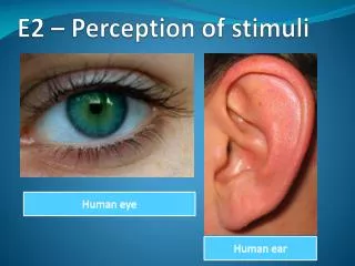
E2 – Perception of stimuli Human eye Human ear
Human sensory receptors • receptors detects the changes in both internal and external environment • they transform the stimuli energy into a nerve impulse that can be sent to the central nervous system (CNS) which in turn coordinates an appropriate response. • receptors are located in the sense organs such as the eye, ear, skin, tongue etc.
Human sensory receptors & stimuli that can detect Type of receptor Stimulus detected Example of receptors mechanoreceptors pressure, sound hair cells in the ear chemoreceptors chemical substances taste buds on the tongue & olfactory cells along the nose thermoreceptors temperature nerve endings in the skin & hypothalamus of the brain photoreceptors light rods and cone in the retinas of the eye hydroreceptors humidity ? nocireceptors pain sensory nerve ending in the skin
Human eye Eye lashes Sclera Pupil Iris Eye lid
Label the diagram of the human eye below sclera pupil choroids retina ciliary body / muscles iris lens cornea fovea blind spot optic nerve
Structure Function(s) tough outer layer of the eye which overs and protect eyeball. Sclera Choroid prevents internal reflection of light and nourish retina. Retina contains rods and cones which convert light into nerve impulses. Ciliary Body a ring of muscle controlling the shape and curvature of the lens. Iris controls the pupil size thus controls entry of light. Pupil a hole in the iris that lets light into the back of the eye. Lens accommodation & focusing of light onto the retina. Cornea bends incoming light focusing it on the retina. Fovea a tiny area of densely packed cones for detailed and coloured vision. exit point of the optic nerve cutting through the retina so no rods or cones Blind Spot Optic Nerve carries the impulses from the rods and cones to the visual center of the brain.
Annotate the diagram of the retina below Axon of the ganglion cell direction of light movement Ganglion cell Bipolar cell Synapse Rod Cone Pigment Sclera
Compare rod and cone cells Rod Cells Cone Cells rod cells more effective in low light intensity cone cells more effective in high light intensity cone cells are sensitive to a specific colours (wavelength) rod cells detect a broad range of colours (wavelength) a single cone cell passes impulses to a single nerve fibre groups of rod cells pass impulses to a single nerve fibre rod cells more sensitive to movement cone cells give higher visual acuity (sharpness ) rod cells respond more slowly to light cone cells respond more rapidly to light rod cells spread through retina cone cells concentrated in centre of retina (at fovea ) rod cells contains one type of pigment (rhodopsin) cone cells contains three types of pigment (iodopsin) rod and cone cells are both are photosensitive
Compare rod and cone cells Rod Cells Cone Cells rod and cone cells are both are photosensitive • cone cells more effective in high light intensity • cone cells are sensitive to a specific colours (wavelength) • a single cone cell passes impulses to a single nerve fibre • cone cells give higher visual acuity (sharpness ) • cone cells respond more rapidly to light • cone cells concentrated in centre of retina (at fovea ) • cone cells contains three types of pigment (iodopsin) • rod cells more effective in low light intensity • rod cells detect a broad range of colours (wavelength) • groups of rod cells pass impulses to a single nerve fibre • rod cells more sensitive to movement • rod cells respond more slowly to light • rod cells spread through retina • rod cells contains one type of pigment (rhodopsin)
Contralateral processing of visual stimuli • rod & cone cells in the retina convert light into nerve impulses • impulses pass to bipolar cells • bipolar cells pass impulses to sensory neurons of the optic nerve • at the optic chiasma, impulses cross over to the opposite optic nerve • impulses continue to the thalamus where optical information is processed • images form in the visual cortex of the brain
Edge enhancement • edge enhancement is a ‘pre- central nervous system ‘processing of information on the retina • it enhances contrast at the edges (boundaries of different objects)and provides more detail to the visual system of the environment • in certain regions of the retina, single ganglion cell receives information form a number of rods and cones, such a region is called receptive field • the fewer the rods and cones that supply a single ganglion the smaller the receptive field & the higher visual acuity i.e. the detailed information one sees
Label the diagram of the ear below Semi circular canals Middle ear bones Oval Window Auditory Nerve Round window Cochlea Eustachian tube Eardrum Auditory canal Pinna
How sound is perceived by the ear • sound waves reaching eardrum cause it to vibrate • vibrations are passed to bones of middle ear which amplify them • the bones pushes the oval window which cause a pressure wave in the fluid-filled cochlea • As the oval window moves in, the round window moves out, this allows the fluid in the cochlea to move freely backward & forward • the vibrations caused by fluid movement pushes the membrane on which the hair cells (mechanoreceptors) sits, triggering nerve impulses in the auditory nerves • the nerve impulse is carried to the auditory cortex in brain through auditory nerve for interpretation