Download
1 / 25
250 likes | 434 Vues
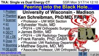
Peering into the Black Hole…. University of Wisconsin - Madison Ken Schreibman, PhD/MD, FACR Professor – UW MSK Section Sylvester Youlo, MD PGY4 – UW Orthopedic Surgery James Brittin, MD PGY4 – UW Radiology Department Frank Ranallo , PhD, DABR Physicist- UW Radiology Department

E N D
Peering into the Black Hole… • University of Wisconsin - Madison • Ken Schreibman, PhD/MD, FACR • Professor – UW MSK Section • Sylvester Youlo, MD • PGY4 – UW Orthopedic Surgery • James Brittin, MD • PGY4 – UW Radiology Department • Frank Ranallo, PhD, DABR • Physicist- UW Radiology Department • Matthew Squire, MD, MS • Associate Professor, UW Orthopedic Surgery
Disclosures Bullet Pts. • Neither I nor my family have any financial disclosures • We are members of the Disney Vacation Club • UW Radiology Department has a partnership with GE • I will try to cover all 10 Bullet Points in less than 10 minutes • No fancy PowerPoint animations in this talk • For fancy PowerPoint see my ePoster… …or visit my website
Peering into the Black Hole… 140kVp Axial • This is the problem • Poor CT visualization inside cupped femoral component • Can’t see bone-metal interface • ? • Can wellsee around tibial tray Lateral Radiograph B,W 57yoM CT: Sagittal
Pig Knee + Human Co-Cr Femoral Component • To simulate the bone-metal interface within the cupped femoral component
ImplantedbySylvesterYoulo,OrthoSurgResident • DrillingTarget • Lesions • Pounding component in place
We Created Target “Erosions” Target 6x6mm 3mm deep Target Target 7x7mm 5mm deep 14x9mm 7mm deep 3D CTwithout metal
Placed in sealable Tupperware™ Filled with water to simulate attenuation of soft tissues surrounding the knee Sealed to eliminate air-fluid level CT Scout
CSC • GE 750 HD + GSI • WIMR • • Scanned the phantom repeatedly • varying one parameter at a time • UW Medical Campus hok.com
“Single Energy” Results: Comparing kV First tested the effect of changing kV using conventional “Single Energy” CT 600 mA, Detail 0.625mm Source image 140 kV 600 mA, Detail 0.625mm Source image 120 kV Target Target 2) 4) For all images: W=3000 L=1000
“Single Energy” Results: Comparing kV 140 kV 120 kV 100 kV Target Target Target • Conclusion: With “SE”CT, use 140 kV 2) 4) 6) For all images: W=3000 L=1000
“Single Energy” Results: Comparing mA • Then, holding kV=140, varied mA 140 kV 600 mA 140 kV 300 mA 140 kV 150 mA Target Target Target • Conclusion:Don’t need to use max mA 2) 8) 10) For all images: W=3000 L=1000
“Single Energy” is actually a Spectrum of Energies • Disclaimer: • On this slide I am going to try to explain physics that my physicist says I don’t understand… • Here’s where things get confusing because there are two types of “voltage”. • The electricity applied into the CT tube is in volts, or kV • The energy of emitted photons in electron-volts, or keV • put kV into CT tube get keV out… • …but what comes out is less than what’s put in! Applying 140kV to the CT tube yields an X-ray spectrum… …nearly all with energies much less than 140keV. • (We can ignore the pointy spikes not as relevant to this discussion) Applying 80kV to the CT tube yields lower energy spectrum. With physics+math, can use the CT attenuation data, measured from these two real energy spectra, to calculate what the attenuation would look like if we had single energy X-rays… Number of X-ray photons emitted from CT tube 140kV 140keV 80kV 0 20 40 60 80 100 120 140 Energy of the X-ray photons (x103 electron-volts) (keV)
“Dual Energy” is actually Single Energy • Conventional “Single Energy” CT generates images from X-rays with a spectrum of energies. • Collecting image data from 2 single energy spectra simultaneously is called “Dual Energy.” • Dual Energy allows generation of images… • …as if the X-rays were of a single energy! Number of X-ray photons emitted from CT tube 140kV 140keV 80kV 0 20 40 60 80 100 120 140 Energy of the X-ray photons (x103 electron-volts) (keV)
GE 750 HD with GSI • GSI: Gemstone Spectral Imaging • What GE calls their DECT • It’s easy to turn on DECT • Click “On” button • With DECT, the presets control the kV and mA
GSI generates bothSE&DE images • Since it takes several minutes to generate DE images, GSI first yields SE Quality Check (QC)images • For technologists to check for coverage, etc. • GE recommends discarding these images. • I found they looked as good as the best SE images we got with high kV and mA. • We archive them. 600 mA SE:140 kV SE:140 kV “600 mA” SECT Acquired earlier GSI: QC Generated automatically Target Target • Conclusion: QC equivalent to SECT 2) 11)
Results: SE&DE CT for Target Lesion 600 mA SE:140 kV “600 mA” DE:140 keV Target Target • Conclusion: DE at least as good as SE 2) 12)
Results: SE&DE CT for Dose • Chart of DLP (Dose Length Product) (mGy-cm) • Values calculated by the CT scanner CHANGING VOLTAGE DECT (keV) Conventional CT (kV) 140 120 100 140 600 696 1202 843 532 CHANGING CURRENT (mA) 300 701 42% lessthan 1202 150 350 • Conclusion:DECT lower dose than SE
Results: SE&DE Metal Blooming (Phantom) 600 mA SE:140 kV “600 mA” DE:140 keV 13mm 10mm • Conclusion:DELessMetalBlooming 2) 12) For all images: W=3000 L=1000
Results: SE&DE Metal Blooming (Patients) SE:140 kV DE:140 keV Can’t see if osteolysis inside femoral component CAN see osteolysis inside femoral component Metal Blooming Much Less Metal Blooming DLP=2764 DLP=1234 55% less than 2764! 630 mA, Detail 3mm Sagittal 600 mA, Detail 3mm Sagittal B,W 57yoM A,C 69yoF
Results: Metal Blooming +/- GE MARs • Metal Artifact Reduction software • Post-processing iterative technique • It can be applied, or not, after scanning • Only available on GE scanners with GSI • “MARs helps significantly in the reduction of artifacts from high density metal implants and allows the accurate visualization of the underlying bone and adjacent soft tissue” DE:140 keV+MARs DE:140 keV 600 mA 600 mA Eliminates Black streak artifacts Black streak artifacts Target Target? www.gehealthcare.com/ct • November 2011 page 29 • Conclusion: MARs may worsen bone-metal interface 13) 12)
Limitations • I’ve only worked with GE 750 HD GSI • That’s what we have at the UW • I’ve only worked with 2 TKA phantoms • Got the same results • Both times failed to see largest target! • Geometry of cupped femoral component? • We weren’t trying to minimize dose • Just wanted to see what advantages DECT offered over SECT
What We’re Presently Doing • Because DECT seems to be no worse than SECT in seeing bone-metal interfaces • SECT & DECT found/missed same targets • and because DECT metal blooming is less… • and because DECT dose is lower… • UW MSK Policy is now this: • For patients getting bone/joint CT scans, • whenever there is metal in scanning FOV, • we recommend using DECT with 140keV. • If DECT not available, use 140kV SECT.
What We’re Planning on Doing • Working with our physicists to optimize the DECT protocols so we can continue to get great images with minimum dose. • Develop body part specific protocols • Hopefully share all of this with all of you at SSR 2015! DE:140 keV 600 mA, Detail 3mm Sagittal A,C 69yoF
Anyone who wants to join me in a bike ride can meet me up front as soon as we’re done