Download
1 / 26
260 likes | 482 Vues
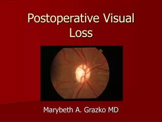
The Americal Society of Anesthesiologists Postoperative Visual Loss Registry. Analysis of 93 spine surgery cases with postoperative visual loss Anesthesiology 2006; 105:652-9 R2 김용일. Postoperative visual loss (POVL). Relatively uncommon but devastating complication

E N D
The Americal Society of Anesthesiologists Postoperative Visual Loss Registry Analysis of 93 spine surgery cases with postoperative visual loss Anesthesiology 2006; 105:652-9 R2 김용일
Postoperative visual loss (POVL) • Relatively uncommon but devastating complication • 0.2-4.5% in spine & cardiac surgery • Ophthalmologic lesions • Ischemic optic neuropathy (ION) • Not consistent with an etiology of globe compression • associated atherosclerotic risk factors • Adverse effects of antihypertensive medications • Sildenafil • Multi-institutional database • Very low number of POVL case • ASA POVL Registry • Established in 1999
Study populations • POVL occurring within 7 days after nonocular surgery • 93 cases associated with spine surgery
Patient and perioperative characteristics • Information collected • Patient demographics • Medical Hx : riskfactors for vascular disease, current medications, surgical history • Obesity, HTN, coronary artery disease, MI, CVA, DM, hypercholesterolemia, tobacco Hx • Intraoperative information • Procedure description, number of levels, type of headrest & surgical frame, position, frequency of eye checks • Duration of anesthetic, surgery, prone positioning • Type of anesthetic, drugs, fluids, estimated blood loss, type of blood products, preoperative & lowest Hb/Hct, urine output • Use of deliberate hypotension, specific hypotensive agents • Intraoperative blood pressure • Presence of hypothermia (<35℃) • Intraoperative events • Cardiiogenic shock, cardiac arrest, seizures, direct trauma to the eye
Ophthalmologic examination characteristics and diagnostic criteria • Ophthalmologic examination • Type of visual deficit, time when visual symptoms were first noted, funduscopic examination, ophthalmologic diagnosis • Classification of specific lesion • Central retinal artery occlusion (CRAO) • Pale ischemic retina with pathognomonic cherry-red spot at macula • And relative afferent pupillary defect or reduced pupillary light reflex • Anterior ischemic optic neuropathy (AION) • Edematous disc with or without peripapillary flame-shaped hemorrhages • And relative afferent pupillary defect or reduced pupillary light reflex • Posterior ischemic optic neuropathy (PION) • Normal early funduscopic examination • With relative afferent pupillary defect or absent pupillary light reflex • Any treatment and prognosis for recovery of vision was noted • Inclusion criteria • Any POVL case associated with spine surgery from ASA POVL Registry • Diagnosis of CRAO, AION, PION, or unspecified ION
Results 93 cases of POVL associated with spine surgery As of June 2005
No statistically significant differences between AION and PION • Demographics, coexisting diseases, surgical characteristics, anesthetic management • Uncertainty whether AION and PION are different disease states with separate etiologies all AION, PION, and unspecified ION were combined under ION
Demographics and coexisting diseases No patient had a preoperative history of glaucoma
Description of operations and positioning (2) All were positioned prone for a portion of procedure Except two anterior spine procedure Eye checks were documented by anesthesiologist in 51%
Anesthetic management Mean anesthetic duration 9.8 ± 3.1 h • General anesthesia • Combination of volatile and narcotic (89%) • Isoflurane (59%), sevoflurane (14%), desflurane (22%), nitrous oxide (29%) • TIVA with propofol and narcotic (2%) • Unknown general anesthetic agents (8%) Median EBL 2.0 L
Anesthetic management (2) Colloid (hydroxydthyl starch or albumin) used in 30% Nadir Hct of 30% or greater in 17% of cases Urine output was less than 0.5ml/kg/h In 24% of cases
Anesthetic management (3) Labetalol or esmolol (n=10) Volatile agents (n=5) Phenylephrine administered in 27% Hypothermia in 10%
Ophthalmologic findings Complete blindness with loss of light perception 64 of 138 affected eyes (47 Pts) Median onset time of reporting Postoperatively 15 h
Spine surgery cases with CRAO Not significantly different Horseshoe headrests in 3 cases Foam pads in 2 cases Miscellaneous headrests in 5 cases Not significantly different Eye checks in 6 cases
Limitation of this study • POVL is low-incidence complication prospective data collection was impractical • Incidence of any POVL cannot be ascertained • Reporting bias, error from retrospective data collection • Increase in POVL in spine surgery may be related to • Increased awareness of the problem • Increased rates of spinal fusion operations
Etiology of ION • Etiology of ION remains unknown • Male patients is 72% • 48% male : 52% female in spinal fusion procedures • National inpatient sample data for 1999 • Influence of sex on ulnar nerve injuries : 70% male • Previous study of ulnar neuropathy • Anatomical differences & hormonal differences • Protective effect of estrogen on cerebral ischemia • Experimental animal models
Etiology of ION (2) • Age • Older patients may be more vulnerable • Young age did not immune to this complication • “Normal” anatomical or physiologic variation in optic nerve blood supply may place more at risk than others • Preoperative identification of high risk group not currently possible
Etiology of ION (3) • Mayfield pins used in 16 pts • With eyes free of pressure ION occurs in absence of pressure on globe • Lack of retinal ischemia in ION • ION in both eyes in the majority more consistent with a systemic etiology • 10 Pts of CRAO (result from globe compression) all had unilateral disease usually with ipsilateral periocular trauma
Etiology of ION (4) • Blood pressure management varied widely • Autoregulation of cerebral blood flow has been well demonstrated • Not clear whether optic nerve also has autoregulation • No difference in lowest BP between visual loss and no visual loss • Case-control study by Myers et al. • Anemia • Cannot be discerned by this study • ION occurs in the absence of anemia
Etiology of ION (5) • Estimated blood loss (EBL) and anesthetic duration EBL of 1,000ml or greater in 82% Anesthetic duration of 6 h or longer in 94% • Not yet enough information to confirm a relation
Etiology of ION (6) • Prone position in 72% • Hypothesis • Venous pressure within optic nerve may increased during prone Perhaps due to venous engorgement • Intraocular pressure increased in prone position • Artery on posterior optic nerve are small end-vessel from surrounding pia • blood flow in posterior optic nerve may be susceptible to increased venous pressure • Case reports of ION after radical neck operation with bilateralinternal jugular vein ligation • Increased venous and intracranial pressure • Hypothesis “compartment syndrome of the optic nerve” • Increased venous pressure and interstitial fluid accumulation • Within relatively nondistensible space • Semirigid lamina cribrosa at optic nerve head • Bony optic canal recommended head-up position and colloid-based fluid resuscitation • Its role in prevention of ION remains undetermined
Etiology of ION (7) • ION almost always occurred • Without any accompanying evidence of vascular injury in other critical organs • Such as heart or brain • Optic nerve vasculature may be uniquely vulnerable tohemodynamic perturbations in prone position in some patients
Summary • More than two thirds of ASA POVL Registry • Related to spine surgery in prone position • 89% : ION • Relatively healthy Pts • Wide range of nadir Hct & BP management multifactorial etiology • EBL > 1,000ml or anesthetic duration > 6 hr 96% • For lengthy spine surgery in prone position risk of visual loss should be considered