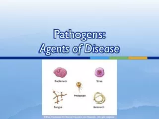
Types of pathogens
Types of pathogens. Pathogens. May be cellular or non-cellular. Cellular pathogens Also known as pathogenic organisms Include: bacteria, protozoa, oomycetes, fungi, worms and arthropods. Non-cellular pathogens Also known as pathogenic agents Include: viruses, viroids and prions. Bacteria.

Types of pathogens
E N D
Presentation Transcript
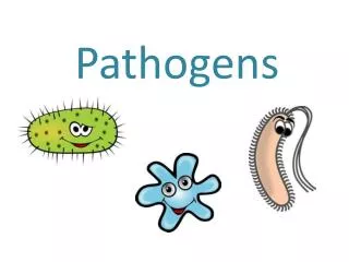
Pathogens • May be cellular or non-cellular. • Cellular pathogens • Also known as pathogenic organisms • Include: bacteria, protozoa, oomycetes, fungi, worms and arthropods. • Non-cellular pathogens • Also known as pathogenic agents • Include: viruses, viroids and prions.
Bacteria • Prokaryotes – no membrane bound nucleus or organelles. • Bacteria have a cell wall and a single major chromosome — a circular thread of DNA double helix. • Replicate by binary fission (20min). • Some bacterial diseases that affect people are diphtheria, food poisoning, wound infections, tetanus, pneumonia, tuberculosis, meningitis, gas gangrene, typhoid fever, gonorrhoea and syphilis.
Classification of bacteria • Bacteria can be classified into different groups. • They are classified on the basis of a number of physical and metabolic characteristics.
Physical characteristics of bacteria Shape • Bacteria can have three basic shapes — round, rod and spiral shapes. • A round-shaped bacterium is called a coccus. • A rod-shaped bacterium is called a bacillus. • A spiral-shaped bacterium is called a spirochaete.
Physical characteristics of bacteria Organisation • Although bacteria are single-celled organisms, they often cluster together in special ways that are used as a basis for classification. • Single, in pairs (diplo), in chains (strepto), clustered (like grapes – staphylo)
Physical characteristics of bacteria Structures • Some bacteria have flagella — thin appendages that originate just below the bacterial wall and are visible with a light microscope when special stains are used. Flagella allow a bacterium to move. Bacteria without flagella cannot move — they are said to be non-motile. • Many bacteria have a layer called a capsuleoutside the cell wall. A capsule is made of slimy gelatinous material and is important in determining the virulence of the bacterium. The virulenceof a bacterium is the degree to which it can cause disease. • Some bacteria, particularly members of the genera Bacillus and Clostridium, form spores. A sporeis a special reproductive structure formed within a bacterial cell. Spores are particularly resistant to heat and drying out. • The cell wallof a bacterial cell is a firm, flexible layer that maintains the shape of the cell and protects the underlying protoplasm. Using the Gram stain, bacteria can be divided into two distinct groups on the basis of a fundamental difference in the chemistry and structure of their cell walls.
Gram stains • Developed in 1884, by the Danish bacteriologist, Joachim Gram (1853–1938). • Gram-positive bacteria stain purple and their cell wall is a relatively thick layer of peptidoglycans, a macromolecule found only in bacteria. • They are generally more susceptible to penicillin and sulfonamide drugs. • Gram-negative bacteria stain pink and have a much more complex, multilayered cell wall, including a layer of peptidoglycans and an additional outer membrane layer of lipids. • The outer layer of lipid compounds enables these bacteria to resist penicillin and other drugs. It also makes phagocytosis of the bacteria very difficult. Drugs such as streptomycin, chloramphenicol and tetracycline are active against Gram-negative bacteria. • The Gram stain is a particularly important stain used to identify the bacteria that are causing an infection because it gives an indication of what drugs will be more effective in the treatment of a patient.
Metabolic characteristics of bacteria Gaseous requirements • Most bacteria are aerobic— they grow and reproduce in the presence of oxygen. • Some bacteria are anaerobicand can live in the absence of oxygen. • Facultative anaerobescan survive whether or not oxygen is present. • Obligate anaerobesgrow and reproduce only in the absence of oxygen. Nutritional patterns • Some bacteria are photosynthetic— they use light as their energy source. Only some of these bacteria are able to use carbon dioxide as their carbon source. • Chemosyntheticorganisms obtain their energy from oxidation reactions. • Some chemosynthetic bacteria can oxidise only organic compounds for their carbon source. Others can oxidise inorganic substances such as ammonia, sulfides and iron compounds and use carbon dioxide as a source of carbon. • Almost all pathogenic bacteria use organic compounds as their source of energy and matter.
Examples of Bacterial Disease • Cocci • Staphylococcus aureus causes skin and wound infections • Streptococcus causes sore throat • Bacilli • Diphtheria – throat infection caused by Corynebacterium diphtheriae • Tuberculosis – lung infection caused by Mycobacterium tuberculosis • Leprosy – skin infection caused by Mycobacterium leprae • Spirochetes • Syphilis and Lyme disease are both caused by spirochetes
Protozoa • Unicellular eukaryotic organisms. • Can reproduce sexually and asexually. • Three classes of protozoans have members that are pathogenic to animals: • Flagellates • Sporozoans • Sarcodinians (amoebas)
ProtozoaN Diseases Flagellates • Trypanosoma – causes sleeping sickness • Giardia – causesdiarrhoea Sporozoans • Plasmodium – causes malaria Sarcodinians, (amoebas) • Entamoeba histolytica – causes amoebic dysentry
Malaria: caused by Plasmodium • The adult stage occurs in humans (the primary host). • Plasmodium larvae migrate to liver and multiply asexually for two weeks. They leave the liver and pass into the bloodstream an infect red blood cells. • Larvae leave the liver and infect red blood cells. There they grow and split into many tiny larvae (merozoites). • After a time, male and female gametes are also formed in the red blood cells and are released when red blood cells burst and can be taken up with blood by the intermediate host, the Anopheles mosquito, is feeding. • Gametes fertilise in the stomach of the female mosquito and develop into larvae which migrate through the stomach wall and into the salivary gland where they undergo asexual reproduction.
Oomycetes • The oomycetes were originally classified as fungi but are now considered part of the kingdom Protista. • They have motile cells (flagella), walls of cellulose and many cellular processes not found in fungi. • Cause diseases such as blight and down mildew on plants. • About 35 species which infect crops including potato, tomato, apple, tobacco and citrus fruits. • Phytophthora cinnammi has destroyed much of the eucalypt timberland in Australia. Its spores can survive for years in moist soil, and are attracted to the roots of the plants they infect by a chemical released from the roots.
Oomycetes - Phytophthora • When Phytophthora spores land on leaf they may be carried by water droplets to other leaves, swim to a germination site, or germinate directly, sending out hyphae that branch out and invade plant tissue • Branching hyphae (haustoria) penetrate living cells and absorb nutrients or release enzymes that digest cytoplasm into molecules that can be absorbed.
Fungi • Fungi can be unicellular or multicellular. They consist of eukaryotic cells with cell walls composed of chitin. • Fungi are important pathogens of plants, causing diseases such as rusts, smuts, ergot and Dutch elm disease. • Fungi that are pathogenic to humans fall into three main groups: moulds, which are filamentous; true yeasts, which are unicellular; and fungi-like yeasts, which are like yeasts but may form long non-branching filaments. • Moulds are multicellular fungi which invade tissue using hyphae while yeasts are unicelluar and reproduce by budding.
Fungal Diseases • Fungal diseases include: • Ringworm (mould) • Athelete’s foot (mould) • Thrush (fungi-like yeast – Candida albicans) • Aspergillus infection (life-threatening for immunocompromised patients). • Some fungi produce toxins that are poisonous to humans, for example Aspergillus species produce toxins (aflatoxins) that are carcinogenic (cause cancer). The fungus grows on peanuts and many grain foods. • Other fungal products such as cyclosporine and penicillin have become important tools in medicine.
Worms (helminthes) • Multicellular, eukaryotic, specialized for the parasitic way of life. • Mouthparts are often modified to form hooks, digestive systems are simple, numerous offspring are produced. • Worms can be divided into two groups: Platyhelminths and Nematodes. • Both animals and plants can be infected with helminthes.
Platyhelminths • Are parasitic flat worms, and include tapeworms, hookworms and blood flukes. • The blood fluke Schistosoma and the hydatid tapeworm are both examples of disease-causing platyhelminths that utilise an intermediate host. • An intermediate host is the host in which larval or juvenile forms of a parasite exist. The primary host is the organisms in which a parasite lives its adult phase. • Tapeworms are the most highly specialized parasitic flatworms. • They have a head that attaches to the wall of the gut. • A neck region, which is the region of growth, • And a chain of segments (protglottids) which each contain male and female reproductive organs. • After fertilization each segment matures into a bag of eggs that breaks off and passes out with the faeces.
Nematodes • Include roundworms, hookworms, and threadworms or pinworms. • Nematodes are the most numerous multicellular animals on earth. There are nearly 20,000 described species classified in the phylum. • Nematodes have been characterized as a tube within a tube. The outside tube is the body wall which consists of muscle layers that are used as a protective covering. The inside tube is the digestive system. • Nematodes range in size from 0.3mm to over 8 metres. • Diseases of humans caused by nematodes are: • Trichinosis – often fatal disease in which worms invade muscle tissue. Caused by eating uncooked, infected pork. • Elephantiasis – swelling of tissue caused by blockage of lymph nodes by adult worms of the Wuchereria bancrofit species. • Nematodes are also important pathogens of plants. They mainly attack roots.
Arthropods • Arthropods (insects) have been associated with many serious diseases. • In most cases they act vectors of disease in plants and animals (they carry pathogens to the host), e.g. • fleas carry bacteria Yersinia pestis (cause of bubonic plague) • Specific mosquitoes carry Plasmodium (cause of malaria) • A few species of insects are parasitic on mammals and actually cause disease or at least discomfort e.g. head lice, body lice, crab louse, fleas and ticks. • Parasitic insects such as psyllids induce the formation of galls (swollen areas) on plant leaves. • Tick fever (caused by cattle ticks Boophilus microplus) is an example of arthropods causing disease in animals.
Viruses • Non-cellular agents that infect all types of organisms. • Consist of either DNA or RNA surrounded by a protein coat and perhaps a modified membrane envelope. • Obligate intracellular parasite – cannot replicate outside of cells. • Interaction between a virus and host cell is specific. • Instructions carried by viruses direct the production of viral proteins and nucleic acid to be assembled into new virus particles. • In most DNA and RNA viruses this process is similar to normal protein production, however the group of viruses known as retroviruses first produce DNA from viral RNA. HIV is an example of a retrovirus. • Some viruses exit the cell by lysis of the cell. • Enveloped virus particles are released slowly by budding from the cell membrane.
HIV • Attachment – virus binds to surface molecule (CD4) of T cell or macrophage • Fusion – viral envelope fuses with cell membrane releasing contents into cell • Reverse transcription – viral RNA is converted into DNA • Integration – viral DNA is inserted into host chromosome (integrated DNA known as provirus and may stay latent for years) • Replication – viral DNA is transcribed and RNA is translated to make viral proteins. Viral genome is replicated • Assembly – new viruses are made • Release – new viruses bud through cell membrane
Viruses Animal Viruses • Associated with a wide range of diseases • DNA viruses (may be double or single stranded) • Smallpox, cowpox, herpes, warts, common cause of sore throats • RNA viruses (usually single stranded) • Polio, hepatitis, influenza, AIDS, Ebola, measles, mumps • Some viruses appear to be able to cause normal cells to become cancerous e.g. hepatitis B virus (liver cancer), Epstein-Barr virus (Burkitt’s lymphoma and nasopharyngeal carcinoma). Plant Viruses • Divided into three main types each of which take their name from the symptoms they produce: • Yellow viruses • Mosaic viruses • Necrotic viruses
Viroids • Tiny circular single-stranded RNA molecules. • About 1/10 the size of smallest virus. • Have no protein coat or membrane envelope. • Infect susceptible cells and replicate themselves. • Only been associated with plant diseases in crops such as potatoes, citrus and coconut.
Prions • A group of abnormal infectious proteins that cause degenerative neurological diseases. • Prions are pathogenic variants of proteins that are naturally produced in nerve cells and certain other cells. The normal "healthy" prions are referred to as PrPc (Prion Protein cellular). • When a defective prion comes in contact with a PrPc (healthy prion) it converts the normal protein into a prion protein. This is the equivalent of the prion replicating itself. • Prions eventually cause a cell to burst and are free to infect other cells. The bursting of nerve cells results in the holes seen in infected brains.
Prion infections Humans might be infected by prions in 2 ways: • Acquired infection (diet and following medical procedures such as surgery, growth hormone injections, corneal transplants) i.e. infectious agent implicated. • Apparent hereditary mendelian transmission where it is an autosomal and dominant trait. This is not consistent with an infectious agent. • No treatment is available for individuals infected with abnormal prions. • Prions are extremely resistant to heat and chemical agents. • Human Prion Diseases • Creutzfeldt-Jakob Disease (CJD) • Variant Creutzfeldt-Jakob Disease (vCJD) • Gerstmann-Straussler-Scheinker Syndrome • Fatal Familial Insomina • Kuru • Animal Prion Diseases • Bovine Spongiform Encephalopathy (BSE) • Chronic Wasting Disease (CWD) • Scrapie • Transmissible mink encephalopathy • Feline sponigform encephalopathy • Ungulate spongiform encephalopathy