Download
1 / 1
10 likes | 158 Vues
Photophysical Studies of Pyrene in Nafion ®. Aaron Fleishman, Mark Flickinger , Tim Roberts, & Evan Spivack Department of Chemistry, The Pennsylvania State University. Abstract. Results and Discussion. Embedding Pyrene in Nafion

E N D
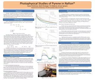
Photophysical Studies of Pyrene in Nafion® Aaron Fleishman, Mark Flickinger, Tim Roberts, & Evan Spivack Department of Chemistry, The Pennsylvania State University Abstract Results and Discussion Embedding Pyrene in Nafion Cyclohexane was found to be an inadequate solvent because it resulted in no detectable pyrene becoming embedded within the Nafion pores. This was confirmed using UV spectroscopy of the Nafion sample. This was expected as cyclohexane is a nonpolar solvent, and both pyrene and water within the pores of swollen Nafion were polar. Ethanol was found to be a very suitable solvent as the initial concentration of the pyrene within Nafion was far greater than the UV spectrophotometer was capable of measuring. The solution was diluted to ___M and the resulting Nafion samples contained a more appropriate concentration of pyrene. This was expected because ethanol is a slightly polar solvent and entropy of mixing caused ethanol with pyrene dissolved in it to mix with water within the pores of Nafion. Pyrene Properties Using Luminescence Spectroscopy The luminescence decay spectrum obtained from laser photolysis showed that pyrene formed both a monomer and an excimer when excited by the laser. This was evidenced by the fact that the intensity versus time data in Figure 2 showed typical monomer behavior using the 415nm bandpass filter, and Figure 3 showed a “build-up” when observed using the 500nm high pass filter. Pyrene monomer emits light in the 415 nm range and pyrene excimer emits light in the 500nm range. When the sample was kept under vacuum for 30 minutes, the ethanol within the pores evaporated and pyrene was observed to form pyrene ground state complex (Py2) . This was determined from the change in the excimer luminescence data. Pyrene was also found to interact with the surface of the Nafion pore as the monomer luminescence data deviated from typical exponential decay. Homemade Spectrophotometer Construction A preliminary absorbance spectrum was obtained from irradiated, 77K Nafion sample, as seen in Figure 4. This spectrum was obtained using a 350nm high pass filter in order to precisely excite the pyrene cations and/or anions. This figure reveals that the spectrophotometer has the potential to analyze samples in the 77K range and would be an adequate detector in further research about the characteristics of radioloysis of Nafion. A procedure, using ethanol as the solvent, was developed for embedding high concentrations of pyrene in Nafion. Attempts at using cyclohexane as the solvent were deemed to be unsuccessful. The properties of pyrene were assessed using luminescent spectroscopy. After the evaporation of ethanol, the existence of a ground state Py2 complex was concluded using reaction kinetics. A cost-effective homemade spectrometer was constructed capable of rapidly collecting data within the range of 300-450 nm, and 350-900 nm, both at 77 K. Introduction Nafion , a perfluorosulfonate ion exchange membrane, has a complex structure that has been studied in a variety of experiments to determine its various uses. Figure 1 below shows the general chemical structure of Nafion : Figure 1: Chemical Structure of Nafion. The sulfonate head groups provide swelling and ion exchange capabilities, while the fluorocarbon backbone stabilizes the membrane thermally, chemically, and structurally. As an ionomer, the polar bonds in Nafion make the membrane very hydrophilic. Water within the membrane tends to cluster and swell around the sulfonate head groups. Although the membrane tends to resist non-polar molecules, the entropy of mixing with water-miscible compounds can be used to drive the incorporation of less polar solvents and solutes into the polar membrane. In this experiment, ethanol was used in this manner to incorporate pyrene into the Nafion membrane. This pyrene served the dual purpose of both revealing the mobility of pyrene molecules in the membrane and the mobility of electron-hole pairs in the membrane produced by gamma radiation. The mobility of pyrene molecules can be gauged by studying the formation of pyrene excimers during photolysis. Excimer formation and dissociation(Eq. 1) is one radiative decay process of interest, another being fluorescence(Eq.2), where excited state pyrene relaxes to its ground state. Eq. 1 Eq. 2 The mobility of electron hole-pairs can be ascertained from the presence of pyrene cations and anions, as the electron and electron holes react with nearby pyrene molecules. These hole-pairs are not stable at room temperature, but can be analyzed at 77K, which necessitates the use of a spectrometer capable of obtaining measurements at this temperature. For this purpose, a spectrometer was custom built and was functional with samples at this temperature. This experiment was only successful in studying the mobility of pyrene in Nafion membrane, although significant conclusions were established with respect to the study of electron hole-pair mobility. Figure 2: Monomer fluorescence decay of pyrene embedded in Nafion using 415nm bandpass filter Conclusion Emission spectra of pyrene monomers in Nafion reveal oxygen, and perhaps many other ethanol soluble gases, to be mobile within the pores of the membrane. This is verified by the dynamic quenching shown by the lifetime changes in the previously mentioned emission spectra. This spectra also reveals the possibility of incorporating non-polar compounds, pyrene in this case, into the polar membrane. The change in lifetime in vacuum pumped samples suggest pyrene relaxation occurring in the absence of ethanol-on the surface of the membrane. This suggests that the experiment successfully adsorbed pyrene on to the polar membrane surface. A similar process could be used to dope Nafion with any number of non-polar compounds. Our analysis of the emission spectra of pyrene excimers indicate two separate radiative decay mechanisms that depend on the concentration of pyrene. Higher concentrations of pyrene, such as in samples that were vacuum pumped, show no time delay in the emission of excimers due to the presence of pyrene dimers which are then excited to form excimers. These results highlight how sensitive concentration is to a kinetic preference for certain photochemical reaction mechanisms. Lastly, the spectrum of 77K Nafion, although deficient in tangible chemical information, proves our success in recording an absorption spectrum at 77K. Had a more properly prepared sample been measured, the spectrometer would have revealed the presence of pyrene cation and anions, and hence the mobility of electron-hole pairs. This indicates that any spectrometer built in this fashion would be capable of obtaining photophysical data of any processes occurring at these low temperatures. Many other physical phenomena which are otherwise thermodynamically unstable may be prepared and analyzed at these temperatures using this custom built spectrometer. Figure 3:Excimer fluorescence decay of pyrene embedded in Nafion using 500nm high pass filter. Experimental Procedure Embedding Pyrene in Nafion at High Concentration Nafion was cut into 4.5 x 4.5 cm squares and soaked in concentrated nitric acid while stirring for 24 hours. The pieces were then soaked in 80% nitric acid for one hour, then in 60% nitric acid for an hour, and finally 40% nitric acid for one hour. This soaking procedure allowed the pores within the membrane to swell and fill with nitric acid solution. Distilled water was used to rinse the Nafion samples in preparation for doping. Pyrene solutions were prepared using cyclohexane and ethanol as solvents, and Nafion was soaked in the solutions for 20-30 minutes in an attempt to embed the pyrene within the pores. Pyrene Properties Using Luminescence Spectroscopy Once a suitable method for embedding pyrene within Nafion was determined, the luminescent properties of pyrene within the Nafion were observed using laser photolysis. A 415 ±10nm band pass filter was used to isolate the monomer specific fluorescence decay data, and a 500nm high-pass filter was used to isolate the excimer fluorescence decay data. An absorbance spectrum was also taken of the pyrene in nafion samples using the Ocean Optics detector and a 350nm high pass filter. Homemade Spectrophotometer Construction A spectrophotometer was constructed using parts found in the local auto supply store to create a rapid data acquisition system to observe the emission spectrum of an irradiated Nafion sample in the 300-450nm and the 350-900nm ranges. The device was constructed using a 100W halogen H3 bulb (Rally Manufacturing Inc., Part # 3125) wired to an EverStart MAXX 12V battery (125 Amp Hours, Part # MAXX-29). A blue (350nm) high pass filter was attached to a clamp in front of the light source. A series of wooden blocks were assembled to hold the sample in place and to block ambient light from the USB UV detector (Ocean Optics Inc., USB 4000) The spectrum readings were recorded using the Ocean Optics software (SpectraSuite). A picture of the setup appears in Figure 5. Figure 4: Absorbance of pyrene in irradiated Nafion at 77 K. References 1Lee, Plato C., and Dan Meisel. "Photophysical Studies of Pyrene Incorporated In Nafion Membranes." Photochemistry and Photobiology 41 (1985): 21-26. Acknowledgements We would like to thank Dr. Bratoljub Milosavljevic for assisting in the experimental procedure and analysis. We would also like to extend our thanks to Ms. Candice Davison for irradiating our samples as well as teaching assistant Matthew Ross for his guidance throughout the experiment. Figure 5: UV Spectrometer Apparatus


