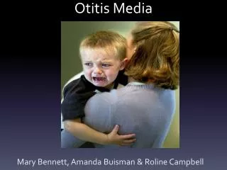
Otitis Media
Otitis Media. 5 y/o Female Incomplete cleft of secondary palate Pain in left ear Tubes 4 years ago. No Medications Cleft has been repaired in 2001 and has healed well. OM Case 1. Otoscopy. Right ear canal clear TM intact Amber colored fluid behind left TM. Tymps. SRT/WR. Audio.


Otitis Media
E N D
Presentation Transcript
5 y/o Female Incomplete cleft of secondary palate Pain in left ear Tubes 4 years ago No Medications Cleft has been repaired in 2001 and has healed well. OM Case 1
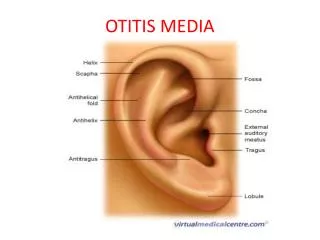
Otoscopy • Right ear canal clear TM intact • Amber colored fluid behind left TM
Diagnosis • Left otitis media
Case 12 History • 9 year old male • Chronic otitis media • Eustachian tube dysfunction • History of PE tube placement
Case 3 History • 2 year old female • Pressure Equalization Tubes placed one month previous for chronic otitis media.
Case 4 History • 3 year old female • Diagnosed with obstructive sleep apnea • Has constant nasal drainage and complains of headaches
Otoscopy • Left tympanic membrane is dull with possible effusion • Right tympanic membrane is intact, fluid bubbles are noted
Recommendation • Myringotomy and tympanostomy tube placement bilaterally • Possible adenoidectomy to correct apnea
Case 5 History • 6 year old male • History of chronic otitis media with effusion, tube placement approximately one month prior
Case 6 History • 23 year old female • Complains of left ear progressive hearing loss • Tympanostomy tubes at age 5 and perforation repair at age 7. • Exploratory surgery to rule out cholesteatoma found only tympanosclerosis Dec of 2000
Otoscopy • Left tympanic membrane bulging with white opacity behind it – no appreciable landmarks, suspicious for cholesteatoma • Right canal obstructed by cerumen which was removed, canal is narrow and collapsible and there is evidence of tympanosclerosis.
Tymps Tymps
CT Scan • Some thickening of the right tympanic membrane • On the left – abnormal tissue filled the external auditory canal associated with calcification or possible reactive bony changes – middle ear appears normal • Left findings are most consistent with keratosis obturans.
Case 7 History • 58 year-old male • Worker’s compensation injury to right ear while using a cutting torch (1 month prior) • A hot slay landed in his ear canal • Developed otorrhea and was given drops • Reported that he could taste the drops (it can be assumed there was a perforation) • All has healed and he feels there has been an improvement in his hearing.
Otoscopy • Small amount of debris in right ear which was removed • Both ear canals are intact and translucent.
Case 8 History • 4 year old male with history of recurrent acute otitis media and chronic otitis media with effusion • Tubes were placed bilaterally approximately 11 months ago
Case 9 History • 4 year old boy • History of right cholesteatoma • Underwent right tympanoplasty 7 months prior • Has has persistent chronic right suppurative otitis media with foul smelling discharge
Otoscopy • Debris was cleaned form the right ear canal and partially removed cerumen filling the left canal but was discontinued due to patient cooperation • Tympanostomy tube still in place in the left TM