Download
1 / 47
480 likes | 539 Vues
This tutorial covers the physiology and pathology of heart failure, nursing interventions, and factors influencing its development such as aging, stress, inflammation, and genetics. Navigate through slides to learn about cardiac physiology, causes of heart failure, important nursing interventions, and more. Test your knowledge on cardiac function, valves, the cardiac cycle, and cardiac output. Explore how the heart pumps blood, its rate, and factors influencing cardiac output. Enhance your understanding of heart failure management strategies.

E N D
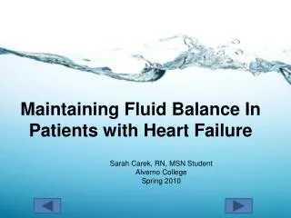
Maintaining Fluid Balance In Patients with Heart Failure Sarah Carek, RN, MSN Student Alverno College Spring 2010
What should I know by the end of this tutorial? • Understand the basic physiology of the heart. • Develop a clear picture as to how Heart Failure is a pathological process. • Recognize how excess fluid volume can result from Heart Failure. • Apply nursing interventions to patients with Heart Failure. • Examine how aging, stress, inflammation and genetics play in the development of Heart Failure.
Tutorial Navigation • Click to advance to the next slide. • Click to go to the previous slide • All buttons will be on the bottom of each page. • Begin the tutorial by clicking on the first topic on the home page.
Slides with Animation • FYI..many slides contain animation. Keep clicking the mouse to advance through the entire animation. If you hover your cursor over areas and it changes from an arrow to a hand…click and animation will occur. • Any underlined words have definitions available. Hover your cursor over the word and the definition will appear.
Home Page Cardiac Physiology Pathophysiology of Heart Failure Causes of Heart Failure Important Nursing Interventions Fresh and New: Aging, Stress, Inflammation & Genetics References Contact Information
CardiacPhysiology Microsoft Clipart (manually altered)
Let’s see what you know already! Aorta Pulmonary Arteries Superior Vena Cava Pulmonary Veins Left Atrium Right Atrium Hover your mouse cursor over each section of the heart until the cursor turns from an arrow to a hand. Then click to reveal its label. **If the mouse has not turned from an arrow to a hand..Do Not Click*** Left Ventricle Right Ventricle Inferior Vena Cava Microsoft Clipart (manually altered)
What do the areas of the heart do?Click on the boxes to the left to find their function. Pumps deoxygenated blood from the body into the right ventricle. Right Atrium Right Ventricle Pumps deoxygenated blood to the lungs to get oxygenated. Left Atrium Receives oxygenated blood from the lungs and pumps it to the left ventricle. Left Ventricle Pumps oxygenated blood to the body to get used for energy
NormalBlood Flow Microsoft Clipart (manually altered)
What about the valves? Pulmonic Valve The valves of the heart assure that blood flows in the right direction. Aortic Valve Mitral (Bicuspid) Valve Tricuspid Valve Click on the diagram to find the four valves. They are half-moon shaped. Again when the cursor turns into a hand, it means you can click on it. Microsoft Clipart (manually altered)
The Cardiac Cycle Click on the boxes on the left to find their definition. Microsoft Clipart Microsoft Clipart (manually altered) Systole The period during the cardiac cycle where the ventricles are contracting and moving the blood forward. Diastole The period during the cardiac cycle where the ventricles are relaxed and filling with new blood. (Porth & Matfin, 2009)
The Heart as a Pump Heart Rate • Cardiac output measures how well the heart is doing its job of pumping blood to the body. • The heart has the ability to adjust cardiac output based on the body’s needs (i.e. exercise, sleep, illness). This depends on preload, afterload, cardiac contractility and heart rate. • Average cardiac output = 3.5 – 8 Liters/minute Cardiac Ouput Stroke Volume (Porth & Matfin, 2009)
Keep clicking to advance through the animation of the slide. Starling’s Law FYI: The fibers only stretch so far. There is a maximum force of contraction that can be achieved. End-Diastolic Volume Stretching of Cardiac Muscle Force of Contraction The more blood in the heart at the end of diastole… The more the cardiac muscle fibers stretch… The greater the force of the contraction. (Porth & Matfin, 2009)
Let’s Review What area of the heart is known as the “power horse” as it needs to pump blood to the entire body? Try Again! Excellent! Right Atrium Left Ventricle Close! No…but you are on the right track. Left Atrium Right Ventricle
What is the function of the cardiac valves? Try Again! Nope. To prevent blood from moving forward. To let air escape preventing an air embolus. You’re Right! Keep Trying! To keep blood moving forward. To prevent blood from leaking out of the heart.
Cardiac Output (CO) is used to measure the efficiency of the heart as a pump. What is the equation used to express CO? A. CO = HR x AV Incorrect B. CO = SV x HR Good Job! C. CO = AV x SV Try Again! D. CO = HR x EF Nope
As the needs of the body change, the heart’s ability to increase cardiac output necessarily needs to change. This ability of the heart depends on what factors? (Choose all that apply.) A. Blood Viscosity Blood Viscosity does not help the heart increase cardiac output. B. Cardiac Contractility Correct! In order to increase cardiac output the heart must be able to change the force of contraction. C. Heart Rate Yes! The heart can change its rate in order to increase or decrease cardiac output. D. Preload Good Job! According to Starling’s Law, if End-Diastolic Volume (preload) increases, the force of contraction will as well. E. Afterload Excellent! Cardiac output can be adjusted based on systemic arterial pressure (afterload).
Pathophysiology of heart failure Microsoft Clipart (manually altered)
Cardiac Function is Impaired HR CO SV Heart Failure happens when there is one or more alterations in preload, afterload or cardiac contractility leading to decreased Cardiac Output. Remember the heart is a pump… • Cardiac Output is a reflection of how well the heart is doing its job of being a pump. • HR either speeds up or slows down depending of what the sympathetic or parasympathetic nervous system tells the heart what to do. • SV is determined by our friends preload, afterload and cardiac contractility. CONTRACTILITY Decreased ATP production and availability of Calcium ions causes the heart to become less able to contract effectively AFTERLOAD If vascular resistance is elevated, the heart has to pump harder to overcome it, putting stress on it which can wear it out Preload A stiffer heart will decrease the amount of blood at the end of diastole (Porth & Matfin, 2009)
These Malfunctions can be Classified into Systolic and Diastolic Dysfunction Click on each box to find the definition. Remember the arrow should turn to a hand before you click. The ventricles are unable to relax and expand, leading to a decrease in preload, stroke volume and cardiac output. DIASTOLIC DYSFUNCTION Impaired contractility leads to a decrease in Ejection Fraction and Cardiac Output. Preload, ventricular wall dilation and pressure subsequently increase. SYSTOLIC DYSFUNCTION (Porth & Matfin, 2009)
Heart Failure can also be classified as either Left Sided Heart Failure or Right Sided Heart failure.
Are you starting to see how excess fluid balance contributes to heart failure? Right Heart Failure Left Heart Failure Blood cannot reach the lungs to get oxygenated. Oxygenized blood from the lungs cannot get to the body. Blood begins to pool in the venous system and tissues. Cardiac Output decreases and blood pools in the lungs. Dependent Edema Jugular Vein Distension Ascites Pooling of Blood in GI Tract Pooling of Blood in Hepatic Veins Decrease in Tissue Perfusion Activity Intolerance Cough, Orthopnea Hypoxia, PND Pumonary Edema (Porth & Matfin, 2009)
Before learning about what causes these malfunctions to occur….Let’s Review Heart Failure can result when one or more of which components of Cardiac Output is impaired? Yes! Good Job. Excellent! Afterload Preload That’s Right! Try Again. This is the thickness of the blood. Cardiac Contractility Blood Viscosity
If the heart was unable to contract to the best of its ability, resulting in an Ejection Fraction of 40%, creating ventricular wall dilation, what type of dysfunction would it be? Try Again! Sorry I made this one up! Diastolic Dysfunction Priastolic Dysfunction No. We didn’t talk about this one. Great! Right Ventricular Dysfunction Systolic Dysfuction
Causes of heart failure ***Remember to place your cursor over the underline words to get the definition*** Microsoft Clipart (manually altered)
Acute Coronary Syndrome Blood begins to pool leading to fluid volume overload!!! Myocardial Infarction and Unstable Angina Decreased blood flow to the myocardium caused by a clot in the coronary arteries. Myocardial Damage (Infarction): evidenced by serum cardiac markers (CK-MB, Troponin). Ventricular Remodeling: the area of the ventricle that is damaged undergoes changes in size, shape and thickness (hypertrophy and dilation). Damage to the ventricle can lead to alterations in preload, afterload and cardiac contractility leading to a decrease in cardiac output. (Porth & Matfin, 2009)
Ischemic Heart Disease Blood begins to pool leading to fluid volume overload!!! Coronary Artery Disease Decreased blood flow to the myocardium from the coronary arteries caused by plaque buildup. Myocardial Damage (Infarction): evidenced by serum cardiac markers (CK-MB, Troponin). Ventricular Remodeling: the area of the ventricle that is damaged undergoes changes in size, shape and thickness (hypertrophy and dilation). Damage to the ventricle can lead to alterations in preload, afterload and cardiac contractility leading to a decrease in cardiac output. (Porth & Matfin, 2009)
Cardiomyopathy Blood begins to pool leading to fluid volume overload!!! Hypertrophic Cardiomyopathy Left Ventricle thickens through genetic predisposition. The heart is unable to fill properly during diastole – Altered Preload. Stroke volume is decreased. Cardiac Output is decreased. (Porth & Matfin, 2009)
Cardiomyopathy cont… Blood begins to pool leading to fluid volume overload!!! Dilated Cardiomyopathy The ventricle is enlarged and wall thickness is decreased due to genetic predisposition, infection, alcohol or unknown cause. Preload and pressure increase. Cardiac Output decreases. (Porth & Matfin, 2009)
Valvular Heart Disease Blood begins to pool leading to fluid volume overload!!! Mitral Valve Disorders Stenosis Regurgitation Because the valve is unable to open fully, the left atrium becomes distended leading to impaired filling during diastole. This leads to decreased cardiac output. Because the valve does not open and close completely, it becomes leaky. Stroke volume is reduced leading to decreased cardiac output. (Porth & Matfin, 2009)
Valvular Heart Disease Cont… Blood begins to pool leading to fluid volume overload!!! Aortic Valve Disorder Stenosis Regurgitation Because the valve is unable to open fully, blood is unable to exit the left ventricle properly and begins to pool. This decreases cardiac output. Because the valve allows blood to flow back into the left ventricle during diastole, cardiac output is decreased. (Porth & Matfin, 2009)
Let’s see if you understand causes of heart failure… Which condition below can lead to acute heart failure? Try Again! This is a cause of chronic heart failure. Excellent! Ischemia to the heart can cause it to not pump blood like it should resulting in heart failure. Dilated Cardiomyopathy Myocardial Infarction Nope. Keep thinking! This is a chronic cause of HF. This is a cause of chronic heart failure. Aortic Stenosis Mitral Valve Prolapse
How does a leaky or malfunctioning valve lead to fluid volume overload (heart failure)? Now you’re thinking! Yay! Blood cannot fill properly during diastole. Cardiac Output is decreased. Now that’s just crazy talk. Good Job! Blood will spill out of the heart everywhere. Blood pools creating edema.
Microsoft Clipart Nursing Interventions
Let’s take a look at how you fit into taking care of heart failure patients… Take a minute to think of nursing interventions you do on a daily basis that might apply to a patient with heart failure.
Fluid Volume Excess Keep clicking to advance the animation. (Ackley & Ladwig, 2006) Things You Would Find On Assessment • Assessment • Lung sounds • Daily weights • Vital signs • I & O’s • Behavior • Drug side effects • Interventions • Restricted sodium diet • Fluid restrictions • Diuretics • Turning patients with edema • Promoting positive self image • Consult with physician Crackles/Wheezes Cyanosis Fatigue Cough Cool Extremities Shortness of Breath Edema Jugular Vein Distention Orthopnea Diaphoresis
Heart Failure and Aging Microsoft Clipart
The Stats • ¾ of the 5 million Americans suffering with heart failure are over the age of 65 and ½ are over the age of 75. • Heart failure is the leading cause of hospitalization among the elderly. • 1 million older adults are hospitalized annually with heart failure. You can see that older adults with heart failure are a huge population for us as healthcare providers. As America ages, the population will only grow larger. (AHA, 2010)
Changes Related to Aging • Arteries stiffen creating resistance against which the heart has to pump. • Heart muscle stiffens, creating filling difficulty. • Cardiac Output declines due to a decline in the maximum rate the heart can reach. • The aged are less able to increase the force of their contractions as is needed during stress, illness and exercise. (Thomas & Rich, 2006)
Remember Starling’s Law… With age the heart muscles stiffens, limiting the amount of blood that can fill during diastole. Thus the fibers cannot stretch as far, creating a decreased force of contraction. End-Diastolic Volume Stretching of Cardiac Muscle Force of Contraction The more blood in the heart at the end of diastole… The more the cardiac muscle fibers stretch… The greater the force of the contraction. (Thomas & Rich, 2006)
Heart Failure and Stress Microsoft Clipart
Remember the equation for cardiac output… In a diseased heart, cardiac output would not increase as it should during the normal stress response. The heart would not be capable of pumping blood efficiently. Therefore a prolonged stress response would only exacerbate the signs and symptoms of heart failure. HR CO SV If the heart rate is under sympathetic nervous system (SNS) and parasympathetic nervous system (PSNS) control….think about what chronic stress would do to the heart rate and subsequently cardiac output…. Stress Release of Epinephrine and Norepinephrine Activation of SNS Increased heart rate and blood pressure (Porth & Matfin, 2009)
Heart Failure and Inflammation Blood begins to pool leading to fluid volume overload!!! • Inflammatory mediators (such as nitric oxide) are activated in a patient with heart failure in an effort to improve cardiac function. • These mediators can damage the endothelium in blood vessels supplying the heart with blood. • Ventricular and vascular remodeling of the myocardium may be a result of this damage. • Ventricular remodeling of the myocardium can result in heart failure because the ventricular cannot pump blood efficiently. (Brunini, et.al., 2009)
Genetics and Heart Failure Blood begins to pool leading to fluid volume overload!!! • Some patients are believed to be at high risk for heart failure due to their genetic make-up. • In a small population, mutations have been found in single genes that trigger the development of heart failure. • Gene mutations have been found in the people with ventricular remodeling and cardiomyopathies • Some mutation examples are • Genes encoding for protein components of the sarcomere which leads to hypertrophy of the myocardium. • Gene mutations resulting in altered dystrophin which normally gives stability to the sarcomere. (Morita, Seidman & Seidman, 2005)
References Ackley, B.J. & Ladwig, G.B (2006). Nursing diagnosis handbook: a guide to planning care (7th Ed.). St. Louis, MO: Mosby Elsevier. American Heart Association (2010). Statistics retrieved from http://www.americanheart.org/presenter.jhtml?identifier=1200026. Brunini, T.M., Mann, G.E., Matsuura, C., Meirelles, L.R., Menden-Ribeiro,A.C., & Moss, M.B. (2009). The role of exercise on l-arginine nitric oxide pathway in chronic heart failure. The Open Biochemistry Journal, 3, 55-65. Morita, H., Seidman, C.E., & Seidman, J. (2005). Genetic causes of human heart failure. The Journal of Clinical Investigation, 115(3), 518-526. Porth, C.M. & Matfin, G. (2009). Pathophysiology: concepts of altered health status (8th Ed.). Philadelphia, PA: Lippincott Williams & Wilkins. Thomas, S. & Rich, M.W. (2006). Heart failure in older people. Generations (Fall 2006), pp. 25-32.
Contact Information Questions, Comments, Concerns? Sarah Carek, RN, MSN student careksl@yahoo.com (608) 577-7866