Download

1 / 17
310 likes | 1.64k Vues
Preparation and Staining of Specimens. Often specimens must be fixed and stained to enhance their visibility and to distinguish morphological properties. Fixation. Preservation of internal and external features of cells Cellular enzymes are inactivated Cell structures are hardened

E N D
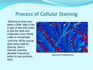
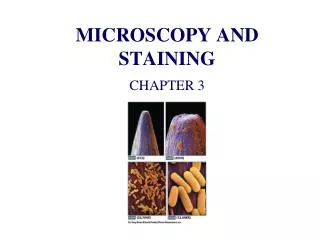
Often specimens must be fixed and stained to enhance their visibility and to distinguish morphological properties
Fixation • Preservation of internal and external features of cells • Cellular enzymes are inactivated • Cell structures are hardened • Organism dies AND adheres strongly to the glass slide
Two types of fixation: • Heat fixing: flame heating bacterial film, preservation of morphology but NOT internal structures • Chemical fixing: chemical fixatives penetrate cells and preserves intracellular components • Acetone • Ethanol • Acetic acid • Mercuric chloride • Formaldehyde • Glutaraldehyde
Dyes and Simple Staining • Dyes contain chromophore groups and bind to cells by ionic, covalent and hydrophobic forces • Basic dyes (positively charged) bind to negatively charged groups • Work best at high pH • Acidic dyes (negatively charged) bind to positively charged groups • Work best at low pH
Simple staining: • One reagent • Usually involves basic dyes • Crystal violet • Methylene blue • Carbol fuchsin • Differential staining: • Used to differentiate groups on bacteria • Examples: • Gram stain • Acid fast stain
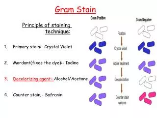
Gram Stain • Gram-positive bacteria retain the crystal violet • Gram-negative bacteria become colorless • Developed by Christian Gram in 1884
Steps: • 1. Crystal violet, 30”, rinse 5” • 2. Gram’s Iodine (mordant that strengthens the association of crystal violet with the cell wall), 1’, rinse 5” • 3. Decolorization: 95% ethanol or isopropanol:acetone, 15-30”, rinse 5” • 4. Counterstain: Saffranin, 1’, rinse 5”, Gram-negative bacteria become pink/red, Gram-positive remain purple
Gram staining is the most widely used differential staining procedure because it divides bacterial species into two roughly equal groups - gram positive and gram negative
Acid-Fast Staining • Some organisms do not stain well with conventional dyes (e.g. Mycobacterium) • Harsher treatment required • Steps: • 1. Heating with basic fuchsin and phenol (Ziehl-Neelson technique) – 5’, cool, rinse 30” • 2. Decolorize: Acid/Alcohol 10-30”, rinse 30” • 3. Counterstain with methylene blue, 2 ‘, rinse 30” • Acid-fast cells remain red due to high mycolic acid content (lipid) • Non-acid-fast cells = blue (counterstain)
Acid-fast staining is a differential staining procedure that identifies two medically important species of bacteria - • Mycobacterium tuberculosis, the causative agent of tuberculosis, and • Mycobacterium leprae, the causative agent of leprosy (Hansen’s disease)
Negative staining • Widely used to visualize diffuse capsules surrounding the bacteria; those capsules are unstained by the procedure and appear colorless against a stained background • Morphology of cells determined • No heat treatment or harsh chemicals • Film spread, stained and dried • Heating would cause shrinkage – distortion of cellular morphology
Deposits generated around cell or dark background observed • Dyes (acidic stain – does not penetrate cells) • Nigrosin • India ink • Eosin blue
Spore staining • Detects endospores generated by Bacillus and Clostridium • Formed within the cell dormant • Hardy in adverse conditions • Various sizes, shapes and locations • Steps (Schaffer-Fulton procedure): • Heat + malachite green • Rinse • Counterstain with saffranin
Capsule stain • Gently – no heat fixation or may get shrinkage • Steps: • Grow culture in skim milk broth • Crystal violet • Copper sulfate counterstain
Flagella stain • Flagella are very difficult to observe directly because of their size • Thickness is increased using mordants • Tannic acid • Potassium alum • Stained with Pararosaniline (Leifson method) or basic fuchsin (Gray method)