Download
1 / 12
120 likes | 341 Vues
Learn about basal cell carcinoma, squamous cell carcinoma, sebaceous gland carcinoma, and eyelash abnormalities. Knowledge of symptoms, causes, treatments, and distinguishing features highlighted.

E N D
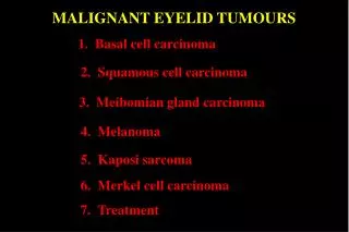
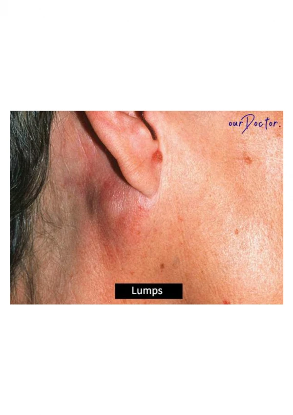
Basal cell carinoma Squamous cell carcinoma Sebaceous gland carcinoma Please notice in the upcoming pictures that Loss of the eyelashes in the vicinity of the tumor always suggests malignancy.
Basal cell carinoma • The most common malignant tumor of the eye lids. • Mainly in the lower lid. • characteristics: • -painless lesion • - slowly growing • - locally invasive • - without metastasis • - can be: nodular, sclerosing • or ulcerative (rodent ulcer). • treatment: • Complete excision • radiotherapy • - Cryotherapy – less effective • prognosis: • Very good but deep invasive tumors are difficult to treat.
Squamous cell carcinoma Rare Age older than 50 years More in Males Uv light is a risk factor for both Squamous cell Basal cell carcinoma. also more common on the lower eyelid characteristics: - Hard nodule or sclary patch. - Locally invasive - Metastasis - They can arise de novo or from pre-malignant lesions . Treatment - Complete Excision
Sebaceous gland carcinoma • very Rare … 0.8% of all lid tumors • Carcinoma of the meibomian and Zeis glands • typically found in women, more often in the seventh decade of life . • usually are on the upper lid margin • Characteristics : • - Highly invasive • - metastasize • may mimic either a recurrent • chalazion or chronic blepharitis • Treatment • - by surgical excision . • Lymph node evaluation is • necessary to evaluate metastasis. • Prognosis • is good with no metastasis, However, sebaceous lesions have a high incidence of recurrence and metastasis
This Sebaceous gland carcinoma … masquerading as Blepharoconjunctivitis. - don’t forget the loss of eyelashes indicates malignancy !
Abnormalities of the lashes Trichiasis Distichiasis
Trichiasis a common condition Where the eyelashes will be directed backward towards the glob , against the cornea It’s distinct from entropion. Complicated by corneal abrasion Symptoms : The eye becomes red and irritated , foreign body sensation, tearing , sensitivity and sometimes pain when exposed to light
Causes : • - Infectious : Trachoma, Herpes zoster • - Autoimmune ,Inflammatory • - Postsurgical • Lower lid transconjunctival approach for floor fracture repair or blepharoplasty • After ectropion repair • - Chemical • Alkali burns to the eye • Medical drops (eg, glaucoma drops) • -Thermal burns to face/lids • treatment: • Epilation of the affected eyelashes with electrolysis, cryotherapy . • An underling abnormal lid position is treated surgically
Distichiasis is a rare disorder defined as the abnormal growth of lashes from the orifices of the meibomian glands on the posterior lamella of the tarsal plate Two types : acquired and congenital. In the acquired form, most cases involve the lower lids. Lashes can be fully formed or very fine, pigmented or nonpigmented , properly oriented or misdirected. The congenital form is autosomal dominant with complete penetrance. It can be isolated or associated with ptosis, strabismus, congenital heart defect, or mandibulofacial dysostosis. This defect may be related to the epithelial germ cells failure to differentiate completely to meibomian glands, instead they become pilosebaceous units, pilo = hair.
Thank you Instructor : Dr. Arkam By Sofian bni awwad