Download
1 / 30
300 likes | 821 Vues
Preterm diagnosis of genetic skin diseases. Genodermatosis. Group of diseases which are due to genetically determined disorder.(monogenic abn .) Genetic disorder are often grouped into three categories: Chromosomal [numerical , structural] Single gene

E N D
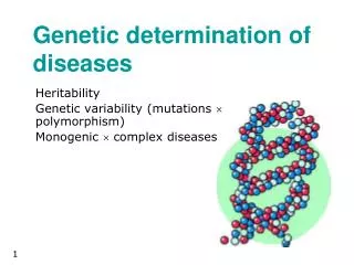
Genodermatosis • Group of diseases which are due to genetically determined disorder.(monogenic abn.) • Genetic disorder are often grouped into three categories: • Chromosomal [numerical , structural] • Single gene • Polygenic [involve complex interaction of genes].
Single gene(mendelian inheritance) which seen in most genodermatosis include: • Aut. Dominant (heterozygous) • Aut.recessive(homozygous) • X-link recessive • X-link dominant
Prenatal diagnosis: is the detection or exclusion of a heriditary disease in utero.divided into: • Invasive include: • DNA based method ( CVS , AS ) • fetoscop and fetal skin biopsy • preimplantation genetic diagnosis B. Non-invasive :1. detection of fetal cells in the maternal blood 2. U/S
DNA- based methodes • Aminocentesis : is a mean of obtaining aminiotic fluid and its cells for morphological, cytogenic, biochemical or molecular analysis • Aminiotic fluid cells are derived from fetal epidermis, alimentary , genitourinary mucosa and amnion • Began in the late 1960s and early 1970s , usually performed at about 17weeks(16-20) where there is adequate fluids volume to minimize the chance of hitting the fetus.
Risk 0f aminocentesis:1.pregnancy loss (especially if done befor 14 weeks) • 2.Increase risk of malformation • 3.Aminorrhea • Indication:use to asses disease associated with abnormal DNA synthesis and repair,metabolic disorder that result in abn. Protien synthesis
Chorionic villus sampling(CVS) • Chorionic villi are of fetal origin,it’s a useful source of fetal DNA, also began at the end of 1960s, taken at (10-12)weeks, either transcervically or transabdominally depend on position of placenta • Risk of CVS: fetal loss (1-2%) but less than AS • Cong. Anomalies (limb reductin defect) so not recommended befor 10 weeks
Tissue obtained from CVS need to be cleaned to exclude maternal cells then subjected to analysis by polymerase chain reaction(PCR) or biochemical analysis , also can be cultured for diagnostic confirmation of the findings obtained by direct analysis
In preperation for DNA-based prenatal testing DNA samples should be available from both parents & the affected family member to screen for pathogenic mutation & informative genetic markers.
FETOSCOPY &FETAL SKIN BIOPSY (FSB) Fetoscopy involve the insertion of fiberoptic endoscope into the pregnant uterus using sedation and local anasthesia At (16-20)weeks FSB represent the earliest form of prenatal testing for inhereted Skin diseases but this approach has largely been superseded by DNA based methods
FSB was first performed in early 1980s for diagnosis of congenital bullousicthyosiformerythroderma&herlitz JEB. FSB initially performed with the aid of fetoscope to visualize the fetus, with improvement of sonography FSB started to be taken under U/S guidance FSB was examined only by light microscopy & transmission EM. but with development of immunohistochemical tests help to complement ultrastructural analysis in establishing the diagnosis especially in case of EB. FSB also used in icthyosis &oculocutaneous albinism
Side effects of FSB • 1.sampling error • 2.inadequacy of samples for analysis • 3.difficulty in interpreting the immunological &morphological features • 4.postnastal scar are usually incospicuous • 5.fetal loss
Main indication of Fetoscopy • Direct visualization of uterine contents which of particular value in identifying defects of facial features,limbs & digits, head&spines,abd.wall &genetalia • Fetal blood sampling from umbilical cord • Tissue biopsy ex. From liver,skin
Preimplantation genetic diagnosis (PGD) PGD is an alternative method that is able to provide information about genetic status of an embryo, it refers to the removal of a single cell from an embryo generated in vitro for genetic testing to diagnose a recurrent serious heritable condition & thereby to avoid the implantation of affected embryo,
it involves assisted conception techniques to generate embryo in vitro,a single cell is then sampled as tissue representative of the whole embryo&analysed for the presence of specific genetic or chromo. abn
PGD • THE techniqes was developed in UK in late 1980s & was used to avoid transmission of adrenoleucodystrophy & X-linked mental retardation, selection of female embryo to avoid X-linked diseases,, dystrophic EB & junctinal EB • Mainly indicated for couples who find that termination at any stage of pregnancy is un acceptble
PGD In vitro fertilization intracellular Sperm injection Biopsy of a single cell from a blastocyst (3-day-old embryo for PGD
NON-invasive methods • It is the assessment of fetal DNA in the maternal circulation, this involves isolation of fetal cells in the maternal circulation or analysis of free fetal DNA in the maternal plasma • In late 19th century, placentally derived trophoblast were first demonstrated in the pregnant mothers • Several fetal cell populations present in maternal blood(eryth,lymph&troph)
Non-invasive M • Limitation:there is only about one fetal cell per ml of maternal blood (detection difficult), sensitvity rate for fetal cell detection 40% , false + detection rates > 10%, fetal cells persist in maternal circulation for months-years(their detection of dubious value) • Fetal DNA (constitutes 3-6% of cell-free DNA in the maternal plasma,its main source apoptotic fragment of syncytiotrophoblasts, unlike nucleated cells free fetal DNA cleared rapidly (mean ½ life 16 min),it is detcectable as early as 4 week . its conc. Increase with progresses of gestation.
Non-invasive methods(3) • Limitation of fetal free DNA:1. very low concetration, 2.genetic information inherited from father can be targeted, cant used to screen maternal alleles(masking of maternal allele by maternal DNA) • Now fetal DNA used to test for RhD blood group&fetal sex determination in women at high risk for X-link condition.
Fetal U/S u/s image very informative in detection heart,bone or CNS abnormality but gross pathology of most inherited skin disorder is usually too subtle&beyond resolution of this approach • Snow- flake sign in amniotic cavity may be a marker of fetal skin sloughing in certain disorders ex.junctional EB, & harlequin icthyosis
Three-dimentional U/S Improve analysis of morphology &has been used in the prenatal Dx. Of harlequineicthyosis by screening for atypical facial dysmorphism (useful for gross morphological abn.),hypohidroticectodermal dysplasia, linear nevus sebaceous, cutis verticisgyrata, identification of associated non-cutaneous features ex.gastricdiltation&polyhydraminos in junctinal EB with pyloric atresia
At present TD-U/S can only be used as prenatal diagnostic tool after about 18 weeks gestation which is too late to become an acceptable & established method of assessment . So it has limited role in clinical practice regarded inherited skin diseases
Prenatal diagnosis for sever inherited skin disorders: 25 years experience • 269 prenatal DX. Performed since 1979-2004 using variety of approaches (FSB, CVS, PGD) • FSB examined for morphological&immunohistochemicalabn. • DNA-based tests involved screening by nucleotide sequencing, restriction enzyme digests&linkage analysis
RESULTS • OF 269 tests 191 were FSB[138 EB(88 junctional,48 dystrophic),37 icthyosis(22 harliquin),oculocutaneous albinism 12], 76 CVS [75 EB(40 junctional ,35dystrophic), 1 EEC syndrom],PGD for skin fragility-ectodermal dysplasia synd.all tests provided accurate DX.&fetal loss rate was about 1%
Further issues will be raised now that preimplantat- ion diagnosis is feasible for genetic susceptibility to certain disorders usually malignancies that occure in adult life • Psoriasis & atopic dermatitis families with clear single gene causation might also be candidate for preimplantation DX. For genetic susceptiblity in the future