Download
1 / 22
220 likes | 602 Vues
MLAB 1315- Hematology Fall 2007 Keri Brophy-Martinez. Chapter 5: The Erythrocyte. Erythrocyte Maturation. Erythropoiesis Production and maturation of erythrocytes Erythropoietin (EPO) The growth factor that stimulates RBC production

E N D
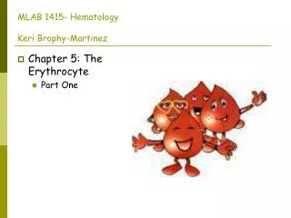
MLAB 1315- HematologyFall 2007Keri Brophy-Martinez • Chapter 5: The Erythrocyte
Erythrocyte Maturation • Erythropoiesis • Production and maturation of erythrocytes • Erythropoietin (EPO) • The growth factor that stimulates RBC production • Released in response to decreased levels of oxygen in the body tissues • Hormone produced and released by the kidneys which acts on committed RBC stem cells to stimulate red cell maturation and release into the blood • With normal levels of EPO stimulation and normal red cell lifespan, about 1% of the red cells in the blood are newly released red cells called reticulocytes. Aged rbc’s are primarily removed by the spleen. • Deficient O2 delivery to the tissues causes the kidney to increase EPO release to accelerate red cell production
Normal RBC in adults • Male 4.7 - 6.1 x 10⁶/µl • Female 4.2 - 5.4 x 10⁶/µl • Infants and children - normals vary by age
Terminology Systems • Normoblast • Rubriblast
Maturation Sequence of Erythrocytes • Stem cell - an unspecified cell that gives rise to a specific specialized cell, such as a blood cell • Multipotential and cannot be identified morphologically • Can self-renew and differentiate • CFU-GEMM: granulocyte, erythrocyte, monocyte, megakaryocyte • BFU-E: burst forming unit • CFU-E: colony forming unit EPO
Maturation Sequence of Erythrocytes • Rubriblast (Pronormoblast) • Size = 14-20 µm • Cytoplasm • Deeply blue (basophilic • Scant amount, may have a perinuclear halo • No granules • Nucleus • Large and round • Reddish-purple with fine chromatin • 1-2 nucleoli (may be bluish) • N:C ratio ( nuclear: cytoplasmic)= 4:1
Maturation Sequence of Erythrocytes • Prorubricyte (basophilic normoblast) • Size = 10-16µm • Cytoplasm • Deeply basophilic indicating RNA activity needed to produce hemoglobin (no hemoglobin is present at this stage) • No granules • Nucleus • Round, large • Chromatin more clumped • No nucleoli • N:C ratio = 4:1
Maturation Sequence of Erythrocytes • Rubricyte (polychromatic normoblast) • Size = 10-12µm • Cytoplasm • Blue-gray to pink-gray (pink indicates that hemoglobin production has begun) • Slight increase in amount • Nucleus • Round and smaller • Chromatin more clumped, irregular • No nucleoli • N:C ratio = 4:1
Maturation Sequence of Erythrocytes • Metarubricyte - Nucleated RBC (orthochromic normoblast) • Size: 8-10 µm • Cytoplasm • Pinker indicating larger amounts of hemoglobin production • Increased amount • Nucleus • Tightly condensed chromatin (pyknotic) • No nucleoli • Mitosis ends at this stage (no more DNA synthesis) • Nucleus is extruded at end of this stage • N:C ratio = 1:1
Maturation Sequence of Erythrocytes • Reticulocyte (diffusely basophilic or polychromatophilic erythrocyte • Size: 8-10µm • Cytoplasm • Diffusely basophilic due to residual RNA • Stain with new methylene blue to see fine reticulum strands • Hemoglobinization is not complete • No nucleus present • Present in circulation for 1-2 days
Lab Methods • New Methylene Blue is asupravital stain it is used to stain reticulocytes. They cannot be identified as reticulocytes from Wright’s stain.
Maturation Sequence of Erythrocytes • Mature erythrocyte • Size = 7-8µm • Volume = 80-100 fL • Cytoplasm • Pink/red • Biconcave shape • Nucleus - none • Present in circulation for about 120 days
Red Blood Cell Membrane • Development • Trilaminar, three-dimensional structure • Outermost layer: glycolipids, glycoproteins • Central layer: cholesterol, phospholipids • Inner layer: cytoskeleton • spectrin • Composed of alpha & beta chains • Join to form a matrix which strengthens the membrane against sheer force and controls biconcave shape • ankrin • membrane proteins
Red Blood Cell Membrane • Function • Shape • Provides the optimum surface to volume ratio for respiratory exchange AND is essential to deformability • Provide deformability, elasticity • Allows for passage through microvessels • Provides permeability • Allows water and electrolytes to exchange • RBC controls volume and H2O content primarily through control of sodium and potassium content
Red Blood Cell Metabolism • Metabolism • These pathways are essential for oxygen transport and maintaining the physical characteristics of the RBC. • Embden-Meyerhof glycolytic pathway • Generates 90% of energy needed by RBC’s • Glucose is metabolized and generates two molecules of ATP (energy). • Hexose monophosphate shunt • Metabolizes 5-10% of glucose. • NADPH is end product • Protects the RBC from oxidative injury. • Most common defect is deficiency of the enzyme glucose-6-phosphate dehydrogenase (G-6PD). • If the pathway is deficient, intracellular oxidants can’t be neutralized and globin denatures then precipitates. The precipitates are referred to as Heinz bodies. (Must use supravital stain to visualize them.)
Red Blood Cell Membrane • Methemoglobin reductase pathway • Maintains iron in the ferrous (Fe2) state. • In the absence of the enzyme (methemoglobin reductase), methemoglobin accumulates and it cannot carry oxygen. • Leubering-Rapaport shunt • Allows the RBC to regulate oxygen transport during conditions of hypoxia or acid-base imbalance. • Permits the accumulation of 2,3-DPG which is essential for maintaining normal oxygen tension, regulating hemoglobin affinity
Checkpoint • Which erythrocyte metabolic pathway is responsible for providing the majority of cellular energy? • For regulating oxygen affinity? • For maintaining hemoglobin in a reduced state?
Checkpoint • Embden-Meyerhof :90-95% • Rapoport-Leubering shunt: oxygen affinity • Hexose-Monophosphate shunt/ Methemoglobin Reductase pathway: iron
Red Blood Cell Metabolism: Summary • Three Areas of RBC metabolism are crucial for RBC survival and function. • RBC membrane • Hemoglobin structure and function • RBC metabolic pathways= cellular energy
Erythrocyte Destruction • Breakdown of the RBC • Toward the end of 120 day life span of the RBC, it begins to break down. This is about 1% of RBC’s per day. • The membrane becomes less flexible. • The concentration of cellular hemoglobin increases. • Enzyme activity, especially glycolysis, diminishes • Aging RBC’s or senescent RBC’s are removed from the circulation by the reticuloendothelial system (RES) which is a system of fixed macrophages. These cells are located all over the body, but those in the spleen are the most efficient at removing old RBC’s.
Extravascular hemolysis • 90% of RBC’s are destroyed extravascularly. • Occurs in splenn, liver and bone marrow • The RES cells lyse the RBC’s and digest them. Components of the RBC are recycled. • Iron is transported by transferrin to the bone marrow to be recycled into hemoglobin. • Amino acids from globin are recycled into new globin chains. • The protoporphyrin ring from heme is broken and converted into biliverdin (green). • Biliverdin is converted to unconjugated bilirubin and carried to the liver by albumin, a plasma protein. • Bilirubin is conjugated in the liver and excreted into the intestine, where intestinal flora convert it to urobilinogen. • Most urobilinogen is excreted in the stool, but some is picked up by the blood and excreted in the urine. • Conjugated (indirect) and unconjugated (direct) bilirubin can be used to monitor hemolysis. • Refer to pg.65
Intravascular hemolysis • 5-10% of RBC’s are destroyed intrasvascularly • RBC breakdown occurs within the blood vessels. • The free hemoglobin α and β dimers that are released into the bloodstream is picked up by a protein carrier called haptoglobin. • The haptoglobin-hemoglobin complex is large and cannot be excreted in the urine. It is carried to the liver where the RES cells and the breakdown process occurs as above. • If there is an increase in intravascular hemolysis, the haptoglobin is used up and free hemoglobin is excreted in the urine (hemoglobinuria). • Free hemoglobin may also be oxidized to methemoglobin which is then broken down extravascularly or to methalbumin which is bound to albumin • Refer to page 66