Download
1 / 28
280 likes | 466 Vues
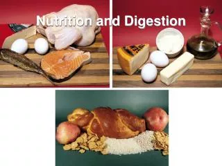
Digestion and Nutrition - 2013 Overall Goal of Digestive Systems A. Obtain organic molecules from environment B. Reduce complex molecules such as proteins, polysaccharides, nucleic acids and lipids to absorbable sizes and

E N D
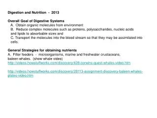
Digestion and Nutrition - 2013 Overall Goal of Digestive Systems A. Obtain organic molecules from environment B. Reduce complex molecules such as proteins, polysaccharides, nucleic acids and lipids to absorbable sizes and C. Transport the molecules into the blood stream so that they may be assimilated into cells. General Strategies for obtaining nutrients A. Filter feeders - microorganisms, marine and freshwater crustaceans, baleen whales. (show whale video) http://videos.howstuffworks.com/discovery/428-corwins-quest-whales-video.htm http://videos.howstuffworks.com/discovery/28113-assignment-discovery-baleen-whales-plates-video.htm
B. Carnivores C. Herbivores 1. the problem with cellulose - requires bacteria or protozoa that produce cellulase 2. pregastric fermentation 3. postgastric fermentation D. Omnivores E. Symbiotic nutrition chloroplasts from algae in the bodies of sea slugs - photo synthetic products feed slugs http://www.newscientist.com/article/dn16124-solarpowered-sea-slug-harnesses-stolen-plant-genes-.html bacteria in digestive tracks of ruminants (cows) http://www.teachersdomain.org/resource/nat08.living.str.living.digest
Perspectives on Digestion • Overview • Mechanical - mastication, stomach, crop, and intestinal motility • Chemical (secretions) - enzymes from saliva, pancreas and intestines • Absorption - intestinal lumen o blood • Assimilation - blood into cells throughout the body. • Motility • Smooth muscle • Pace setter potential - specialized cells in digestive tract produce electric impulses(potentials) that stimulate the gut to contract • Enteric (intrinsic) nervous control (responds to stretch, pH, hormones) • Extrinsic = controlled by central nervous system (brain & spinal cord)
Intrinsic Nerves of the Rat’s Stomach Fig. 14-5, p.619
Ingestion Mouth Receiving Pharynx Foregut Conducting Storage Digestion Crop Motility Digestion (acidic) Midgut (stomach) Secretions Absorption Assimilation Digestion (basic) Hindgut Storage of waste Defecation Fig. 14-1, p.613
The enzyme “cellulase” is required to hydrolyze cellulose. This enzyme is only produced by microorganisms. Animals must provide organs that support the growth of these symbionts. Starch is hydrolyzed to maltose by the enzyme “amylase” which is produced by the salivary glands and the pancreas.
Hydrolysis of a disaccharide (maltose) to two monosaccharides (glucose) Maltase is the enzyme that performs this action. It is produced primarily by the cells that line the intestinal lumen.
Categories of Digestive Enzymes Amylase: starch hydrolysis to maltose Protease: proteins hydrolyzed to peptides Lipase: triglycerides hydrolyzed to fatty acids and glycerol Nucleotidases nucleic acids hydrolyzed to nucleotides
Gastrointestinal Tract (alimentary canal) mouth-anus Accessory Organs Liver Gall Bladder Pancreas
Nasal passages Hard palate Soft palate Uvula Pharynx Epiglottis Esophagus Bolus Trachea Tongue Glottis at entrance of larynx (a) Fig. 14-7a, p.624
Esophagus Fundus Smooth muscle Gastroesophageal sphincter Body Stomach folds Pyloric sphincter Oxyntic mucosa Pyloric gland area Antrum Duodenum Fig. 14-8, p.627
Gastric pit Mucosa Submucosa Table 14-3b, p.632
In oxyntic mucosa Surface epithelial cells Gastric pit Mucous cells Produce pepsinogen Chief cells Produce HCl and Intrinsic Factor Gastric gland Parietal cells Enterochromaffin- Like (ECL) cells Table 14-3c, p.632
Peptic Ulcers • Heliobacter pylori - bacterial infection responsible for 90% • of all peptic ulcers • Treatment with antibiotics
HCl, fats, osmo CCK Secretin
Intestinal Cross Section Layers of the alimentary canal Serosa Smooth muscle cells Muscularis Submucosa Mucosa Microvilli Villus (Villi) Mucosal cells
Negatively charged H2O-soluble portion (a carboxyl group at the end of a glycine or taurine chain) Small lipid (fat) droplet with bile salt molecules adsorbed on its surface Lipid-soluble portion (derived from cholesterol) Large fat droplet Through action of bile salts Lipid emulsion Fig. 14-17, p.644
Lipid Absorption Fig. 14-24a, p.654
Block Diagram of Lipid Absorption Lipids in Duodenum HDL’s and LDL’s re-enter blood and are distributed to other cells throughout the body. Liver takes up Chylomicra and converts them to HDL’s and LDL’s Lipids combine with Bile and form Micelles Lipase begins to convert triglycerides to fatty acids Chylomicra move through lymphatic system and enter the blood at subclavian veins. Micelles taken up by mucosal cells Mucosal cells add proteins and extract fatty acids Lipids enter lymph capillaries as Chylomi ra
Transverse colon Haustra Taeniae coli Descending colon Ascending colon Ileocecal valve Appendix Sigmoid colon Cecum Rectum External anal sphincter (skeletal muscle) Internal anal sphincter (smooth muscle) Anal canal Fig. 14-26, p.657
Rumen Reticulum Omasum Abomasum To small intestine (b) Fig. 14-27b, p.661