THE PROTEASES
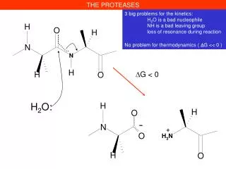
THE PROTEASES. -. +. 3 big problems for the kinetics: H 2 O is a bad nucleophile NH is a bad leaving group loss of resonance during reaction No problem for thermodynamics ( D G << 0 ). D G < 0. H 2 O:. THE PROTEASES. H 2 O.

THE PROTEASES
E N D
Presentation Transcript
THE PROTEASES - + 3 big problems for the kinetics: H2O is a bad nucleophile NH is a bad leaving group loss of resonance during reaction No problem for thermodynamics ( DG << 0 ) DG < 0 H2O:
THE PROTEASES H2O The peptide bond is « stable » under physiological conditions t1/2 102 years despite its thermodynamic instability Hydrolyzed under very harsh conditions, in acid (HCl 6 M, 110°C, 24-72 h) or base (KOH 1 M 100°C 24 h)
THE PROTEASES H2O 3 big problems for the kinetics: H2O is a bad nucleophile NH is a bad leaving group loss of mesomery during reaction 5 biological strategies to solve the problems, 5 classes of proteases 1. Serine proteases Trypsin in mammalian digestion Coagulation factors (Thrombine) 2. Cysteine proteases Papaine Cathepsines (in the lysosomes) 3. Aspartyl proteases Pepsine (in our stomach) AIDS virus protease 4. Metal ion proteases (Zn2+ ) Carboxypeptidases 5. Threonine proteases Proteasome
THE PROTEASES Where the proteases act? Exo-proteases (exo-peptidases, cut amino acids from the N- or from the C-terminal of proteins/peptides) Endo-proteases (cut in the interior of proteins/peptides) Specificity Non-specific (Proteinase K, used for stability studies of proteins) Specific proteases (Trypsine X-X-Arg↓X-X et X-X-Lys↓X-X recognize 1 side chain Very specific (Thrombine LeuValProArg↓GlySer ) recognize 6 side chains
THE PROTEASES IMPORTANT HINT! The protease best substrates are UNFOLDED proteins. Compact protein domain are not hydrolyzed, instead the connecting domains are hydrolyzed Essential application in protein biochemistry and imunology for domain preparation by controlled proteolysis Classical experiment: Porter 1955, preparation of immunoglobulin fragments by treatment with papain or pepsin Disulfide bonds
THE PROTEASES SDS gel electrophoresis
Stanley B. Prusiner infected proteinase K • strange pathogen (resistance to UV, heat etc…) • transmission to mouse (incubation time 150-300 days) • 1975-77: transmission tohamster (70 days) The presence of the Prion protein is demonstrated by the resistence to proteolysis
Blood clotting Blood clotting results from a cascade of reactions. In a cascade, a signal initiates a series of steps, each of them catalyzed by an enzyme. At each step the signal is amplified. In blood clotting the activated form of one clotting factor catalyzes activation of the next. Very small amounts of the initial factors trigger the cascade, => rapid response to trauma (e.g., damage to a blood vessel).
Blood clotting Fibrin fiber
Conversion of fibrinogen to fibrin causes clotting. The final step of clotting is conversion of fibrinogen to fibrin by thrombin, a protease. Fibrinogen has 6 protein chains (2x A, B and ), folded into globular units connected by rods. Thrombin cleaves 4 peptides from the A and B chains in the central globule, resulting in fibrin monomer ()2.
Carboxyl ends of the - and chains interact with the newly exposed N-terminal regions => polymerization (protofibrils).
Blood Coagulation Hemostasis
Fibrils are stabilized by cross-linking: formation of amide bonds between lysine and glutamine by transglutaminase, which is activated from protransglutaminase by thrombin. The network of fibrils forms the clot.
Activation of thrombin. Thrombin activates fibrinogen, but how is thrombin activated ? Thrombin is activated by proteolytic activation of prothrombin with factors Xa (also a protease) and Va. Activation removes a gla and 2 kringle domains. Modular structure of prothrombin
Use of chromogenic substrates for studying the proteases Benzoyl-Phe-Val-Arg ↓ Thrombine (enzyme in blood coagulation) Natural substrate: le fibrinogen (a large protein, about 2000 residues) The product (p-nitro-aniline) est yellow (l 380 nm)
1. The serine proteases N O H2O H O H N O H O H2O Proteases having an essential serine in the active site Protéases Trypsine Chymotrypsine Elastase Subtilisine (Bacilus subtilis) Same mechanism for esterases Lipases Esterases (acétyl)choliesterase Amides and esters have similar structure and reactivity
Identification of active serine in serine proteases An Unusually Reactive Serine in Chymotrypsin Chymotrypsin is inactivated by treatment with diisopropylphosphofluoridate (DIPF), which reacts only with serine 195 among 28 possible serine residues. No reaction with the unfolded enzyme, nor with free serine
Identification of active serine in serine proteases 100 50 0 Percent Inhibition of activity (%) Addition of Substrate protects DIFP Inhibition No substrate + DIFP X Add substrate + DIFP & substrate S Reaction time
Evidence for Histidine Participation inactivator (TPCK) substrate With [14C]TPCK get 1 equiv. [14C] bound; pepsin hydrolysis gives a [14C] peptide with His-57 modified
4 Catalytic elements in serine proteases Specificity pocket Aa 189, 216, 226 Oxyanion hole Aa 193-195 Substrate binding Aa 214-216 Catalytic triad Ser195, His57, Asp102 Chymotrypsin
Chymotrypsin STRUCTURE: David BLOW 1968
: Serine is the NUCLEOPHILE Histidine is a BASE: it binds the serine’s proton and decreases its pKa from 15 to about 7 The aspartate keeps the histidine in the correct orientation (an old theory: proton relay, but the proton does not move)
but identical active site! Subtilisin An example of CONVERGENT evolution Trypsin
Thrombin and Chymotrypsin are HOMOLOGS (Almost identical structures, similar sequences
Evolution is most often DIVERGENT « Ancestral » gene, duplication and separate evolution by mutation Trypsine, chymotrypsine, élastase Structure très similaire Famille de protéines Triade: Ser195, His57, Asp102 Different genes, protein evolution To a similar active site configuration A few examples of CONVERGENT evolution Subtilisine Structure très différente Triade: Ser221, His64, Asp32
Many serine proteases age activated by proteolysis (protection of the cells which synthetize the proteases)
Serine Protease Mechanism - Chymotrypsin Hydrophobic pocket Active site residues
This is a reaction INTERMEDIATE and not a transition state Reaction coordinate
The C-terminal part of the substrate dissociated and leaves the Acyl-enzyme
STEP 2: Acyl-enzyme hydrolysis
Kinetic demonstration of the serine protease mechanism: burst kinetics
Demonstration of the serine protease mechanism: site-directed mutagenesis Nature. 1988, 332(6164):564-8. Substrate: N-succinyl-L-Ala-L-Ala-L-Pro-L-Phe-p-nitroanilide Bacillus amyloliquefaciens subtilisin, these functional elements impart a total rate enhancement of at least 109 to 1010 times the non-enzymatic hydrolysis of amide bonds
Reaction mechanism of a serine protease (in this case, subtilisin) Note the three residues of the “catalytic triad”: Ser221, His64, & Asp32.
Demonstration of the serine protease mechanism: site-directed mutagenesis Subtilisine Km kcat/Km kcat Asp32 H64 S221 (µM) (M-1 s-1) s-1 Asp His Ser 220 250000 55 Ala His Ser 480 5 0.0024 Asp Ala Ser 390 0.1 0.000039 Asp His Ala 420 0.1 0.000042 Ala Ala Ala 420 0.1 0.000042 kSerHisAsp/knon-enzymatique = 3 750 000 000 kAlaAlaAla/knon-enzymatique = 3 000
When very low residual activities are expected, a very low level of contamination with other proteases is a serieus problem. How has this been avoided? Serine24 (on the protein surface) has been replaced by a Cysteine which makes possible protein purification by covalent affinity chromatography. • A second problem could be the mis-incorporation during traduction. An error rate of 1/1000 can be a problem !
DG DG G E + S DG ES E + P Reaction coordinate 3. Ascertaining the role of specific amino acids in catalysis by site-directed mutagenesis can easily by interpreted if the chemical step is rate-limiting (A). If the substrate binding is rate-limiting (B), it is well possible to miss important details of the mechanism. The measured rate is slower with the mutant No apparent effect! B A G E + S DG ES E + P Reaction coordinate Replacing an active-site residue will slown down reaction in A but not in B
Take home lesson: even with no catalytic residues, the enzyme still accelerates the reaction better than 1000-fold the rate of the uncatalyzed reaction.Way to bind that transition state!
Demonstration of the serine protease mechanism: site-directed mutagenesis Site-directed mutagenesis and the role of the oxyanion hole in subtilisin. Bryan P, Pantoliano MW, Quill SG, Hsiao HY, Poulos T. Proc Natl Acad Sci U S A. 1986 Jun;83(11):3743-5. Reaction intermediate is stabilized Asn side-chain in subtilisin: its role CAN be probed by site-directed mutagenesis!!! Reaction intermediate is stabilized by main-chain NH in chymotrypsin: its role cannot be probed by site-directed mutagenesis