PN 132 – Day 2
750 likes | 1.02k Vues
PN 132 – Day 2. Respiratory Nursing - Laboratory and Diagnostic Procedures. Objectives. Identify normal parameters for common diagnostic tests. Describe the purpose, significance of results, and nursing interventions related to diagnostic examinations of the respiratory system.

PN 132 – Day 2
E N D
Presentation Transcript
PN 132 – Day 2 Respiratory Nursing -Laboratory and Diagnostic Procedures
Objectives • Identify normal parameters for common diagnostic tests. • Describe the purpose, significance of results, and nursing interventions related to diagnostic examinations of the respiratory system. • Identify how to interpret ABG lab findings
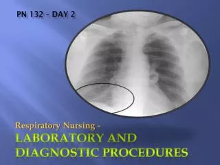
Chest Xray • roentgenogram, chest radiographs • done to diagnose a chest disorder. • -visualizes: • lungs • vertebrae • ribs • heart • clavicles • major thoracic vessels • humeri • scapulae
Nursing Interventions • Pre-exam preparation • Client needs to remove • Articles of clothing • Anything containing metal • Wears a patient gown • Tied (never pinned)
Computed Tomography [CT Scan] • Pictures of small layers of lung tissue • Scanner can rotate at different angles • Non-invasive • Minimal radiation exposure • Painless
Helical CT Scan • Also called • Spiral CT Scan • Volume-Averaging CT Scanning • Continuously obtains images • Produces faster and more accurate results • Images the chest and abdomen in just one breath-hold (about 30 seconds) • Contrast imaging can also be done
Nursing Interventions • Patient teaching re: the procedure • Patient prep similar to CXR • Monitor post-contrast injection • s/sx of allergic reaction to the contrast medium • Allay the anxiety of claustrophobia • Answer questions
Pulmonary Angiography • Radiographic contrast material injected into the pulmonary arteries • Visualization of the pulmonary vasculature • Detects: • Pulmonary Embolism (clot) • Variety of lesions (tissue damage) in the pulmonary vessels
When would we use Pulmonary Angiography ? • Let’s say…Pulmonary Embolism is suspected • A lung CT scan will be done first • If lung CT scan is negative • pulmonary embolism is ruled out • However…. • If the Lung CT Scan is uncertain • If there are 2 or more other possibilities Definitive diagnosis may involve a pulmonary angiography
VENTILATION-PERFUSION SCAN (V/Q Scan) • Studies • Air Flow (ventilation) • Blood Flow (perfusion) • Purpose is to look for blood clots (pulmonary emboli) in the lungs • V/Q used in mathematical equations that calculate airflow and blood flow
V/Q Scan Procedure • Performed in the radiology department • 2-step procedure • Multiple pictures of the chest are taken from different angles • Special camera that detects a radionuclide • For half of these pictures • Patient breathes from a tube that has a mixture of air, oxygen, and a slightly radioactive version of a gas called xenon • Measures airflow in different parts of the lung • For the other half of the pictures • Camera tracks an injected radionuclide • Determines blood flow in different parts of the lung • Embolus is suspected in areas of the lung that have good airflow but poor blood flow. • Except for the minor discomfort of having the IV placed, the test is painless. It usually takes less than one hour.
Pulmonary Function Test [PFT] • Assesses the presence and severity of disease in large and small airways • Composed of various procedures • Lung Volume • Ventilation • Pulmonary Spirometry
PFT • http://www.youtube.com/watch?v=1rjN2_hDXEY
Pulmonary Function Test [PFT] • Lung Volume • Vital Capacity • Volume of air • Maximum inhalation slowly exhaled • Inspiratory Capacity • Largest amount of air • Inhaled in one breath from the resting expiratory level • Total Lung Capacity • Volume of air in the lung after maximum inhalation
Pulmonary Function Test [PFT] • Ventilation • Measures the amount of air inhaled and exhaled in each respiratory cycle • Pulmonary Spirometry • Evaluates the amount of air that can be forcefully exhaled after maximum inhalation
Bronchoscopy • Bronchoscope • Flexible tube with a camera
Bronchoscopy • Bronchoscope is passed through the nose, larynx, trachea, and bronchi • Local or general anesthetic may be given • Observation of the respiratory structures • Tissue samples may be obtained • Secretions can be suctioned for sampling
Bronchoscopy Procedure http://www.youtube.com/watch?v=KqZc1JqArco
Nursing Interventions Pre-Procedure • Explain Procedure • Answer Questions • Allay Anxiety Post-Procedure • NPO until gag reflex returns (about 2 hours) • Semi-Fowler’s position • Turn side to side • Eases removal of secretions • Monitor for laryngeal edema and laryngospasms • Monitor sputum • S/Sx hemorrhage • Blood streaked sputum is expected to last for a few days after biopsy
Sputum Culture Sputum specimen collected - obtained for microscopic studies - culture and sensitivity
Cytology Cell studies - tests for the presence of abnormal or malignant cells - Any body secretions - pleural fluid - abdominal fluid - pericardial fluid, etc. Melanoma
Lung Biopsy Open lung biopsy Transbronchial Done through a bronchoscope Cells and/or secretions obtained for testing Done when pulmonary disease cannot be diagnosed by other means Nursing Interventions Same as for bronchoscopy
Thoracentesis Surgical Perforation Into the pleural space Excess pleural fluid removed Sample obtained for diagnostic testing Instillation of medication into the pleural space
Thoracentesis • Diagnostic tests on pleural fluid - Specific gravity - RBCs - WBCs - protein - glucose - culture and sensitivity - abnormal or malignant cells - biopsy of the pleura.
Nursing Interventions During procedure: Monitor vital signs Monitor respiratory status Monitor general appearance After procedure: Position patient comfortably on the unaffected side Continue to monitor patient respiratory status and vital signs
Pulse Oximetry (SpO2) Continuous peripheral monitoring of the pulse and saturation of oxygen in the blood Assessment of gas exchange Noninvasive method Normal Range = 95-100% Probe is applied to a finger, earlobe, toe, or the bridge of the nose.
Pulse Oximetry (SpO2) Monitor displays the hemoglobin- oxygen saturation and pulse rates Allows the nurse to see continuous changes in the pt.’s oxygen saturations Interventions can happen immediately.
Pulse Oximetry - Precautions For Best Results Do Not: Place the probe on an extremity that has an injury, B/P cuff, IV, or arterial line in place Place the probe over a pulsating vascular blood bed Protect the probe from strong light May affect the reading Remove nail polish May affect the reading Avoid excess patient movement Note: Hypothermia, hypotension, and vasoconstriction can affect readings
Pulse Oximetry - Considerations SpO2 < 85% Weakened ability of hemoglobin to release oxygen to the plasma SpO2 < 70% Life threatening If patient has circulatory problems: Probe may not be able to transmit If Pulse Oximeter unable to accurately transmit: ABGs should be drawn
Understanding ABGs Acid/Base Balance
Arterial Blood Gases [ABG] Done by Arterial Blood Draw Measures lung’s ability to: Oxygenate arterial blood Remove Carbon Dioxide Evaluates Acid-Base Balance
ABG Draw • Procedure: • 5 mL (5cc) syringe with needle • Usually radial artery is used • Approximately 1mL (1cc) of blood is drawn • Apply direct pressure to the site for at least 5 minutes • If patient is taking aspirin, coumadin, or other blood thinner apply direct pressure for 20 minutes
ABG Results • Measures: • pH • Hydrogen (H+) ions present • PaO2 • Partial pressure of oxygen dissolved in arterial blood • HCO3 • Calculated value of amount of bicarbonate (HCO3-) in the bloodstream • PaCO2 • Amount of carbon dioxide dissolved in arterial blood • Base Excess (B.E.) • Amount of excess/insufficient level of bicarbonate in the system • SaO2 • Arterial oxygen saturation
Acid/Base Balance • Body uses Acids and Bases to maintain homeostasis • Respiratory and Renal systems both contribute to the balance • Respiratory System can effect change in 15-30 minutes • Renal System takes hours to days to have an effect • Acids and Bases are counter balanced • ABG measures this acid/base status of the arterial blood
Understanding Acid/Base Balance • Respiratory System • Intake of oxygen and release of CO2 • CO2 is a volatile acid • Decreased respiratory rate = retain CO2 • increased CO2 in blood • Acidosis • Increased respiratory rate = blow off CO2 • decreased CO2 in blood • Alkalosis
Understanding Acid/Base Balance • Renal System • Kidneys get rid of non-volatile acid • (Hydrogen – H+) • Maintain constant Bicarbonate (HC03-)level • Bicarbonate is the body’s base
Understanding Acid/Base Balance “Acidosis” • Excess H+ (Hydrogen) • elevated pH • Decreased HCO3- (Bicarbonate) • Kidneys try to compensate by • Excreting H+ • Retaining HCO3- • Respiratory Systemtries to compensate by • Increasing ventilation • Blow off CO2 to decrease the acidosis
Understanding Acid/Base Balance“Alkalosis” • Decrease of H+ and excess of HCO3- • Kidneys try to compensate by • Excreting HCO3- • Retaining H+ • Respiratory System tries to compensate by • Decreasing ventilations (hypoventilation) • Retain CO2 and decrease the alkalosis
RespiratoryAcidosis When we lose the balance…
RespiratoryAlkalosis When we lose the balance…