Download
1 / 57
580 likes | 927 Vues
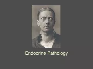
The Endocrine system for dental students DR IBRAHIM HASSAN ALZAHRANI FRCPath -UK Chairman of Pathology Departement Faculty of Medicine . CONTENTS:. Pituitary gland Hypopituitarism Hyperpituitarism Posteroir pituitary syndromes Thyroid galnd Hypothyrodism Hyperthyrodism Goiter

E N D
The Endocrine system for dental studentsDR IBRAHIM HASSAN ALZAHRANI FRCPath -UK Chairman of Pathology DepartementFaculty of Medicine
CONTENTS: • Pituitary gland • Hypopituitarism • Hyperpituitarism • Posteroir pituitary syndromes • Thyroid galnd • Hypothyrodism • Hyperthyrodism • Goiter • Thyrodidtis • Tumors • Parathyroid glands • Hyperparathyroidism • Hypoparathyroidism
Adrenal gland • Cortex • Medulla . Tumors • Multiple endocrine neoplasia • Endocrine pancreas (D.M )
This is the normal appearance of the thyroid gland on the anterior trachea of the neck..
Normal thyroid seen microscopically consists of follicles lined by a cuboidal epithelium and filled with pink, homogenous colloid
Hypothyroidism: • Causes: • structural or functional • 95% are due to: • Surgical or radiation ablation • Hashimoto’s thyroiditis • Primary idiopathic hypothyroidism
Cretinism • This is uncommon disease of childhood due to failure of thyroid to synthesize thyroid hormones hypothyroidism
Neurologic & myxedematous patterns • Clinically: • mental retardation • growth retardation (short stature) • coarse facial features with dry skin and protruding tongue • muscle weakness and umbilical hernia
Myxedema • Hypothyroidism in adult. • - Clinically: • appear insidiously & subtle • lethargy & weakness with slow speech • cold intolerance with cool & rough skin • menstrual problems & psychosis • cardiac changes: cardiac output, hypertrophy, (myxedema heart), pericardial effusion • deposition of mucopolysaccharides in connective tissue • atherosclerosis ( cholesterol)
Hyperthyroidism • Excess thyroid hormone (Thyrotoxicosis) • Causes: • primary diffuse toxic hyperplasia (Grave’s disease) > 95% • toxic multinodular goiter • toxic adenoma • certain form of thyroiditis • secondary to pituitary or hypothalamic lesion
Clinical features: • nervousness and emotional instability • menstrual changes • fine tremors of the hands • heat intolerance with warm skin and sweating • weight loss despite a good appetite
Eye changes: (exopthalmos, widened palpebral fissures, staring gaze) • Cardiac changes: (tachycardia, palpitations, atrial fibrillation and thyrotoxic cardiomyopathy----- cardiac failure) • skeletal muscle atrophy and fatty infiltration • lymphadenopathy • fatty change of the liver • Osteoporosis
Thyrotoxicosis Upper, thyrotoxicosis Lower, after treatment
Goiter • Goiter simply means enlarged thyroid
Diffuse Goiter • Characterized by diffuse symmetrical enlargement of thyroid (200 - 300 gm) with normal thyroid function. • Hypofunction may occur early in the course . • Usually occurs in: Endemic areas ( iodine & goiterogens) or • Sporadic (physiological ,autoimmune , familial ).
Multinodular Goiter • Characterized by nodular asymmetrical enlargement of thyroid (up to 1000 gm) • Slowly evolves from diffuse goiter.It can be toxic or non-toxic
Solitary thyroid nodule • Size (symptoms) • Possible hyperfunction • Usually colloid nodule >70% • Adenoma 20-30% • Carcinoma <5% • - Radioactive iodine (Hot & cold nodule) • FNA & biopsy • Thyroid function
Solitary thyroid nodule • Invisigations: • thyroid hormons: (T3,T4,TSH) • radiological examinations : * ultrasound (cystic/solid) * radioactive iodine (cold/hot) • Fine needle aspiration cytology
GRAVE’S DISEASE • Primary Diffuse Toxic Hyperplasia • The most common cause of thyrotoxicosis • It is an autoimmune disease • Classically shows: • 1-Exopthalmos (proptosis) • 2-Dermopathy (pretibial myxedema) • 3-Hyperthyroidism • Common in ♀ 3rd & 4th decade • ♀ : ♂ = 10 : 1 • HLA – DR3 & Familial predisposition • Other autoimmune diseases may occur
Pathogenesis • B-cells secrete autoantibodies against mainly TSH – Receptors (Abs. against microsomes, thyroglobulin, T3 & T4 can be seen)
Morphology • Gross: diffuse symmetrical enlargement of thyroid
THYROIDITIS • Hashimoto’s thyroiditis • Subacute (granulomatous,DeQuervian) thyroiditis • Chronic lymphocytic (painless) thyroiditis • Riedel’s fibrous thyroiditis
Hashimoto’s thyroiditis • This is an autoimmune most common type of thyroiditis characterized by symmetrical modesty enlarged thyroid responsible for most cases of primary goiterous hypothyroidism.
Pathogenesis • B cells autoantibodies against microsomes and thyroglobulin. • Cell-mediated destruction of the gland • ♀ : ♂ = 10 : 1 middle-aged • Higher incidence of autoimmune disease
Clinical Course • Euthyroid--- hypothyroid • Moderate goiter • Hashitoxicosis(hyperthyroidism) occasionally • 5% - B cell lymphoma or rarely papillary carcinoma of thyroid
THYROID TUMOURS 1-BENIGN: Follicular adenoma 2-MALIGNANT: • Carcinoma of thyroid • Papillary carcinoma • Follicular carcinoma • Medullary carcinoma • Anablastic carcinoma • Lymphoma Others – rare (sq. ca, sarcomas, metastasis)
ADENOMA • Always follicular adenoma • No papillary adenoma of thyroid. • Solitary & encapsulated. • No capsular invasion. • Histology: Follicles –> macro (colloid), micro (fetal), normal size (simple), trabecular (embryonal). • Sometimes composed of Hürthl cells (oncocytic) Hurthle cell adenoma.
CARCINOMA OF THYROID • Causes: • Ionizing radiation • Hashimoto’s thyroiditis • Grave’s disease?
Papillary Carcinoma 60-70% • The most common type • Young age 20-50y , F:M=3:1 • Forming papillae and psammoma bodies • Cells typically show ground-glass appearance with clear grooved nuclei “Orphan Annie” and intranuclear inclusion • 50% at presentation Cervical LN metastasis • Haematogenous spread is rare (not common)
Follicular variant of papillary carcinoma : No papillary formation . The nuclei shows typical nuclear ground glass appearance of papilary crcinoma. • Grow slowly with indolent course • Occult microscopic variant
Follicular Carcinoma • Macroscopically often encapsulated similar to adenoma • Histologically : composed of follicles with no papillary formation and no groundglass nuclear changes. • sometimes the cells are oncocytic (Hurthle cell carcinoma).
Follicular Carcinoma • Haematogenous spread (lung, bone, liver. . ) • Poorer in prognosis than papillary carcinoma. • Represent approximatly 15% • Most patients are >40y • TYPES: 1- minimally invasive FC. 2- widely invasive FC.
Medullary Carcinoma of thyroid <5% • Derived from calcitonin – secreting C-cells • Characterized by formation of amyloid material from calcitonin, surrounded by small to medium sized cells with round to spindle shaped nuclei forming sheets, nests or cords
Medullary Carcinoma amyloid
Medullary Carcinoma • It has slow but progressive growth • Both lymphatic and hematogenous metastasis occurs • 10-20% are familial, multicenteric in young age, associated with MEN 2&3 • Immuno: +ve calcitonin • 80-90% sporadic, solitary, old age
Anablastic carcinoma 5-10% 0ccurs in patient > 60 y • Poorly differentiated, highly malignant tumour usually forms bulky necrotic mass often disseminate extensively through blood • death occurs within 1-2 years (<10% survive for 10y) • Histological variants: • Giant cells, spindle cells(sarcomatoid), squamoid cells
Hyperparathyroidism - Primary Hyperparathyroidism: Increase PTH due to parathyroid lesion (Adenoma/hyperplasia)Hypercalcaemia PTH Hypercalcaemia: • osteoclast to mobilize Ca++ from bone • Ca++reabsorption in the kidney • Ca++ absorption inGit.through vit.D. • excretion of phosphate in urine. • Part of MEN I & II • F : M = 3 : 1 > 40y
Asymptomatic (lethargy&weakness) Bone pain (osteomalacia, osteoporosis & osteitis fibrosa cystica/brown tumor) Renal stones (nephrolithiasis) Nephrocalcinosis Metastatic calcification (blood vessels, soft tissue & & joints) Abdominal pain (peptic ulcer,pancreatitis) and mental change Clinical features
Parathyroid adenoma adenoma normal