Antepartum Hemorrhage (APH)
830 likes | 3.14k Vues
Antepartum Hemorrhage (APH). Saeed Mahmoud, MRCOG,MRCPI,MIOG,MBSCCP Assistant Professor & Consultant Department of Obstetrics & Gynecology College of Medicine King Saud University. Antepartum Hemorrhage. Obstetrics is "bloody business."

Antepartum Hemorrhage (APH)
E N D
Presentation Transcript
AntepartumHemorrhage (APH) Saeed Mahmoud,MRCOG,MRCPI,MIOG,MBSCCP Assistant Professor & Consultant Department of Obstetrics & Gynecology College of Medicine King Saud University
Antepartum Hemorrhage • Obstetrics is "bloody business." • Death from hemorrhage still remains a leading cause of maternal mortality.
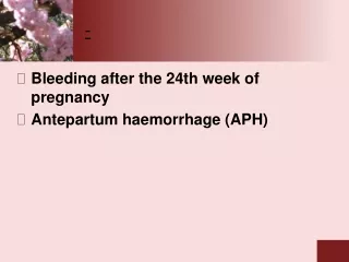
ANTEPARTUM HEMORRHAGE • APH is blood loss Per vagina after 20 weeks’ gestation. • Complicates close to 4% of all pregnancies and is a MEDICAL EMERGENCY! • Is one of the leading causes of antepartum hospitalization, maternal morbidity, and operative intervention.
COMMON CAUSES • Placental Abruption • Placenta Previa • Uterine Rupture • Vasa Previa • Bloody Show • Coagulation Disorder • Hemorrhoids • Vaginal Lesion/Injury • Cervical Lesion/Injury • Neoplasia
Placenta Previa • Defined as a placenta implanted in the lower segment of the uterus, presenting ahead of the leading pole of the fetus. • Total placenta previa. The internal cervical os is covered completely by placenta. • Partial placenta previa. The internal os is partially covered by placenta. • Marginal placenta previa. The edge of the placenta is at the margin of the internal os. • Low-lying placenta. The placenta is implanted in the lower uterine segment such that the placenta edge actually does not reach the internal os but is in close proximity to it.
Placenta Previa • Bleeding results from small disruptions in the placental attachment during normal development and thinning of the lower uterine segment
Placenta Previa • Incidence about 1 in 300 • Perinatal morbidity and mortality are primarily related to the complications of prematurity, because the hemorrhage is maternal.
Placenta Previa • Etiology & Risks • Advancing maternal age • Multiparity • Multifetal gestations • Prior cesarean delivery • Smoking • Prior placenta previa
Placenta Previa • The most characteristic event in placenta previa is recurrent painless hemorrhage. • This usually occurs near the end of or after the second trimester. • The initial bleeding is rarely so profuse as to prove fatal. • It usually ceases spontaneously, only to recur.
Placenta Previa • Placenta previa may be associated with placenta accreta, placenta increta or percreta. • Accreta = adherent to endometrial cavity • Increta = placental tissue invades myometrium • Percreta = placental tissue grows through uterine wall
Placenta Previa • Coagulopathy is rare with placenta previa. • The simplest and safest method of placental localization is provided by transabdominal sonography. • Transvaginalultrasonography has substantively improved diagnostic accuracy of placenta previa. • MRI • At 18 weeks, 5-10% of placentas are low lying. Most ‘migrate’ with development of the lower uterine segment.
Placenta PreviaManagement • Admit to hospital • NO VAGINAL EXAMINATION • IV access • Placental localization
Placenta PreviaManagement Severe bleeding Caesarean section Resuscitate >34/52 Moderate bleeding Gestation <34/52 Unstable Resuscitate Steroids Stable Mild bleeding <36/52 Gestation Conservative care >36/52
Placenta PreviaManagement • Delivery is by Caesarean section • Occasionally Caesarean hysterectomy necessary.
Placental Abruption • Defined as the premature separation of the normally implanted placenta. • Occurs in 1-2% of all pregnancies • Perinatal mortality rate associated with placental abruption was 119 per 1000 births compared with 8.2 per 1000 for all others.
Placental Abruption • external hemorrhage • concealed hemorrhage • Total • Partial
Placental Abruption • What are the risk factors for placental abruption?
Placental Abruption The primary cause of placental abruption is unknown, but there are several associated conditions. • Increased age and parity • Preeclampsia • Chronic hypertension • Preterm ruptured membranes • Multifetal gestation • Hydramnios • Cigarette smoking • Thrombophilias • Cocaine use • Prior abruption • Uterine leiomyoma • External trauma
Placental Abruption • Pathology • Placental abruption is initiated by hemorrhage into the decidua basalis. • The decidua then splits, leaving a thin layer adherent to the myometrium. • development of a decidual hematoma that leads to separation, compression, and the ultimate destruction of the placenta adjacent to it.
Placental Abruption • Bleeding with placental abruption is almost always maternal. • Significant fetal bleeding is more likely to be seen with traumatic abruption. • In this circumstance, fetal bleeding results from a tear or fracture in the placenta rather than from the placental separation itself.
Placental Abruption • The hallmark symptom of placental abruption is painwhich can vary from mild cramping to severe pain. • A firm, tender uterus and a possible sudden increase in fundal height on exam. • The amount of external bleeding may not accurately reflect the amount of blood loss. • Importantly, negative findings with ultrasound examination do not exclude placental abruption. Ultrasound only shows 25% of abruptions.
Placental Abruption • Shock • Consumptive Coagulopathy • Renal Failure • Fetal Death • Couvelaire Uterus
Placental Abruption • Management: Treatment for placental abruption varies depending on gestational age and the status of the mother and fetus. • Admit • History & examination • Assess blood loss • Nearly always more than revealed • IV access, X match, DIC screen • Assess fetal well-being • Placental localization • Delivery
Uterine Rupture • Reported in 0.03-0.08% of all delivering women, but 0.3-1.7% among women with a history of a uterine scar (from a C/S for example) • 13% of all uterine ruptures occur outside the hospital • The most common maternal morbidity is hemorrhage • Fetal morbidity is more common with extrusion
Uterine Rupture • Classic presentation includes vaginal bleeding, pain, cessation of contractions, absence/ deterioration of fetal heart rate, loss of station of the fetal head from the birth canal, easily palpable fetal parts, and profound maternal tachycardia and hypotension. • Patients with a prior uterine scar should be advised to come to the hospital for evaluation of new onset contractions, abdominal pain, or vaginal bleeding.
Uterine Rupture • Multiparity • Non-vertex fetal presentation • Shoulder dystocia • Forceps delivery • Excessive uterine stimulation • Hx of previous C/S • Trauma • Prior rupture • Previous uterine surgery
Uterine Rupture • Management: Emergency laparotomy
VasaPrevia • Rarely reported condition in which the fetal vessels from the placenta cross the entrance to the birth canal. • Incidence varies, but most resources note occurrence in 1:3000 pregnancies. • Associated with a high fetal mortality rate (50-95%) which can be attributed to rapid fetal exsanguination resulting from the vessels tearing during labor
VasaPrevia • There are three causes typically noted for vasa previa: • Bi-lobed placenta • Velamentous insertion of the umbilical cord • Succenturiate (Accessory) lobe
VasaPrevia • Risk Factors: • Bilobed and succenturiate placentas • Velamentous insertion of the cord • Low-lying placenta • Multiple gestation • Pregnancies resulting from in vitro fertilization
VasaPrevia • Management: • When vasa previa is detected prior to labor, the baby has a much greater chance of surviving. • It can be detected during pregnancy with use of transvaginal sonography. • When vasa previa is diagnosed prior to labor, elective caesarian is the delivery method of choice.
Kleihauer-Betke Test • Is a blood test used to measure the amount of fetal hemoglobin transferred from a fetus to the mother's bloodstream. • Used to determine the required dose of Rh immune globulin. • Used for detecting fetal-maternal hemorrhage.
Apt test • The test allows the clinician to determine whether the blood originates from the infant or from the mother. • Place 5 mL water in each of 2 test tubes • To 1 test tube add 5 drops of vaginal blood • To other add 5 drops of maternal (adult) blood • Add 6 drops 10% NaOH to each tube • Observe for 2 minutes • Maternal (adult) blood turns yellow-green-brown; fetal blood stays pink. • If fetal blood, deliver STAT.
Initial management of APH • Kleihauer-Betke test • Apt test • CTG • Observation • Placental localization • Speculum examination when placenta previa excluded • Anti-D if Rh-negative • Admit • History • Examination • NO PV • Nurse on side • IV access/ resuscitate • Clotting screen • Cross match