Download

1 / 46
560 likes | 1.22k Vues
Voltage-gated Sodium Channels. XuelianMa. Introduction. In1952 , Hodgkin and Huxley demonstrated the role of sodium channels in action potential electrogenesis and predicted many of the properties of these channels

E N D
Voltage-gatedSodium Channels XuelianMa
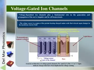
In1952,Hodgkin and Huxley demonstrated the role of sodium channels in action potential electrogenesis and predicted many of the properties of these channels • Sodium channels play central roles in electrogenesis in almost all types of neurons
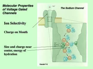
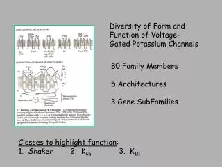
Molecular structure • A large α subunits of 260 kDa and smaller βsubunits of 30–40 kDa
The α subunit • is sufficient for expression of functional sodium channels • The β subunits • modulates the kinetics and voltage dependence of sodium channel activation and inactivation • modulateslocalization of sodium channel.
The β subunits • four NaVβ subunits in total • β1 and β3 are associated non-covalently with α subunits • whereas β2 and β4 form disulfide bonds with α subunits
sodium channel properties are modulated in a cell-type specific manner • native neuronal background G616R variant of NaV1.7
Inherited Erythromelalgia(红斑肢痛症) • L858H NaV1.7 mutation • produce hyperexcitability within DRG neurons and hypoexcitability within sympathetic ganglion neurons. the selective expression of NaV1.8 within DRG neurons, and its absence within sympathetic ganglion neurons
Figure 6. Excitability of sympathetic ganglion neurons is reduced by erythromelalgia NaV1.7 mutation L858H but can be rescued by coexpression of NaV1.8 A, suprathreshold responses recorded from representative superior cervical ganglion (SCG) neurons transf...
Two important principles • The first is that the effects of ion channel mutations on neuronal function are not necessarily unidirectional or predictable on the basis of changes in channel function per se; A single ion channel mutation can have divergent functional effects in different types of neurons. • The second is that cell background and specifically the precise make-up of the electrogenisome can shape the functional effects of an ion channel mutation.
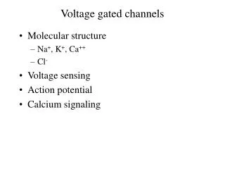
Nav channel neuronal distribution • Sodium channel α subunits are expressed in different excitable tissues (Table1; Goldin, 2001). • NaV1.1, 1.2, 1.3 and 1.6 are the primary sodium channels in the central nervous system. • NaV1.7,1.8 and 1.9 are the primary sodium channels in the peripheral nervous system. • NaV1.4 is the primary sodium channel in skeletal muscle, • NaV1.5 is primary in heart.
Nav1.3 • Nav1.3 voltage-gated sodium channels have been shown to be expressed at increased levels within axotomized dorsal root ganglion (DRG) neurons and within injured axons within neuromas and have been implicated in neuropathic pain.
The more hyperpolarized component of ramp current from Nav1.3 is more likely to be involved in altering threshold. • The more depolarized second component of ramp current may, in contrast, play a role in inter spike interval pacemaking when neurons or their axons are depolarized after injury.
NaV1.7 • NaV1.7 activates in response to small slow depolarizations close to resting potential so as to produce its own depolarization • The ability of NaV1.7 to boost subthreshold stimuli increases the probability of neurons reaching their threshold for firing action potentials. • NaV1.7 is considered to be a threshold channel
Nav1.8 • Nav1.8 is relatively resistant to inactivation by depolarization (Fig. 2A)and recovers rapidly from inactivation. • NaV1.8 thus produces repetitive firing in depolarized DRG neurons • Nav1.8 producing most of the inward current underlying the action potential upstroke during repetitive firing
Nav1.7 and Nav1.8 function in tandem, with Nav1.7 amplifying small depolarizations to bring the cell to threshold, and Nav1.8 producing most of the inward current underlying the action potential upstroke during repetitive firing .
Nav1.9 • NaV1.9, is characterized by very slow activation and inactivation with a large overlap centred near resting potential
Nav1.9 • this channel contributes a sodium conductance at rest that modulates the excitability of DRG neurons
NaV1.7(and NaV1.6 and/or NaV1.9 in some cells) brings the neuron toward threshold (dashed line), NaV1.8 is largely responsible for the overshooting action potential with minor contributions of NaV1.1, NaV1.6 and NaV1.7 to the action potential upstroke. Multiple sodium channel subtypes participate in electrogenesis in small DRG neurons
In 2005 ,Swensen & Bean • Purkinje neurons from NaV1.6−/− mice in which sodium current density is reduced in the long term, where an upregulation of calcium channels maintains excitability at close to its normal level .
The firing properties of most neurons, are usually maintained within a circumscribed range. • A result of homeostatic regulation of ion channel expression, post translational modification, and/or interaction with binding partners or modulators. Changes in expression of channel ‘B’ can compensate for changes in expression of channel ‘A’ to maintain excitability within a particular range
‘Electrogenistat’ These homeostatic regulation of intrinsic neuronal excitability imply a need for an ‘electrogenistat’ within excitable cells
current versus voltage (I-V) curve • A number of important and useful parameters can be readily derived from this plots, including: • reversal potential , • voltage-dependence (rectification), • activation threshold, • as well as overall quality of voltage-clamp
G = I/(Vm – ENa), • where G is conductance, • I is peak inward current, • Vm is the membrane potential step used to elicit the response • ENa is the sodium reversal potential
Voltage-dependence of activation 50mv -60mv -80mv
Voltage-dependence of activation Boltzmann distribution equation: GNa = GNa;max/{1+ exp[ (V1/2 - Vm)/S ]} V1/2 S
Boltzmann distribution equation: GNa = GNa;max/{1+ exp[ (V1/2 - Vm)/S]} GNa is the voltage-dependent sodium conductance, GNa,max is the maximal sodium conductance, • V1 ⁄ 2 is the potential at which activation is half-maximal, • Vm is the membrane potential • k is the slope.
Fast inactivation process 15mv -10mv -50mv -60mv
Boltzmann function: I = Imax / (1 + exp( V1/2 – Vm)/ S ) V1/2 S ** P < 0.01 vs no CRD group
INa = INa;max/{1+ exp[ (V1/2 - Vm)/k]} • where INa,max is the peak sodium current elicited after the most hyperpolarized prepulse, • Vm is the preconditioning pulse potential, • V1 ⁄ 2 is the half maximal sodium current • k is the slope factor.
Steady state inactivation • Inactivation kinetics
The kinetics of recovery from inactivation • Time course of recovery from fast inactivation Double pulse protocol -10mv -60mv 2nA 5ms