Download
1 / 40
400 likes | 733 Vues
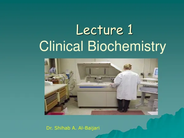
CLINICAL BIOCHEMISTRY 1 ___________________________ School of Biomedical Sciences, Curtin University of Technology. By Mrs Chan Gek Lan & Dr Chan Soo Yin 2 Written Assignments 40% Written Examination 60%

E N D
CLINICAL BIOCHEMISTRY 1___________________________School of Biomedical Sciences, Curtin University of Technology By Mrs Chan Gek Lan & Dr Chan Soo Yin 2 Written Assignments 40% Written Examination 60% Ref: Burtis, CA and Ashwood, ER, Tietz Fundamentals of Clinical Chemistry (4th Edition), WB Saunders 1996. Burtis, CA and Ashwood, ER, Tietz Textbook of Clinical Chemistry (3rd Edition), WB Saunders 1999.
INTRODUCTION________________ ANALYTICAL CHEMISTRY Use of analytical methods & good instrumentation Qualitative & quantitative analyses on body fluids CLINICAL CHEMISTRY Understanding of normal biochemical processes Results to diagnose & treat disease
LECTURE 1 • METHOD EVALUATION • SELECTION & INTERPRETATION • RADIOACTIVITY & ITS MEASUREMENT
1. METHOD EVALUATION___________________________________________________________________________________________________ Method Selection & Evaluation Requirements for Implementation Selecting an Analytical Method Evaluating an Analytical Method Tietz. Fundamentals, Chapters 12
1A. Method Selection & Evaluation • Key steps in implementing new methods • Select new or revised method with care -> Evaluate analytically • Method involves both methodology & instrument Table 13-1: The Keys to Successful Evaluation of a New Method • Apply a clinical perspective for the whole task. • Set a target – an analytical goal • Conduct the right experiments to collect the necessary data. • Use statistical tools properly so that you estimate the errors correctly. • Make objective conclusions
1B. Requirements for Implementation • Practical Requirements – specimen, personnel, cost, quality control, safety • Performance Parameters – properties relating to performance of method • Accuracy – closeness of agreement between measured value of an analyte and its ‘true’ value • Analytical Range – range of concentration or other quantity in the sample over which the method is applicable c. Analytical Sensitivity – quantifies change in signal relative to change in quantity, concentration, or property d. Analytical Specificity – ability of an analytical method to determine exclusively the analyte it measures
Cont. Performance Parameters • Blank Measurements – measurements due to reagent & sample constituents f. Detection Limit – the smallest concentration or quantity of an analyte that can be detected • Interferences – the effect of a compound(s) has on the accuracy of measurement • Precision – the ability to produce the same value for replicate measurements i. Recovery – ability of an analytical method to measure an analyte correctly when a known amount of it is added to samples j . Ruggedness – consistently reliable performance when used by different operators & with different batches of reagents
1C. Selecting an Analytical Method • The principle of the assay • The composition of reagents & reference materials, the quantity provided, & their storage • The stability of regents & reference materials • Possible hazards, appropriate safety precautions • The type, quantity & disposal of waste • Specimen requirements – collection, volume, storage • Anticipated analytical performance – accuracy, precision, range • The reference interval – derivation, values in health & disease • The detailed protocol for performing the test • The availability of technical support, supplies
1D. Evaluating an Analytical Method • Write an evaluation protocol & a method procedure • Document the validity of the method • Determine the reportable range • Determine the components of precision • Determine the accuracy • Estimate the sensitivity • Estimate the detection limit • Test the specificity • Establish the reference interval
2. SELECTION & INTERPRETATION • ___________________________________________________________________________ • Sensitivity & Specificity • Receiver operating characteristic curves • Predictive value Tietz. Fundamentals, Chapter 11
SELECTION & INTERPRETATION • ____________________________________________________________________________________________ • Problem -> clinical tests -> different procedures • Qn: Which procedure to use ? • Evaluate test performance -> select Example: Urologist using Assay to detect prostatic carcinoma Table 12-1: Correct Prediction of Prostatic Carcinoma by Two Prostate-specific Antigen Assays *Does not include patients with benign prostatic hypertrophy Qn: Which test is better?
2A. Sensitivity vs Specificity_________________________________________________ Sensitivity: fraction of those with specific disease that the assay correctly predicts Specificity: fraction of those without the disease that the assay correctly predicts (Table 12-3) Cont. Urologist story: Urologist wants to use assay on BPH patients Benign Prostatic Hyperplasia (noncancerous prostate enlargement) associated with higher PSA If exclude in test -> increase in false-negative If include in test -> increase in false-positive
Table 12-4: Correct Prediction of Prostatic Carcinoma & Benign Prostatic Hyperplasia Fig 12-1: Dot plot of PSA on BPH patients & prostate carcinoma patients • Test A & B are two different cutoffs Lower cutoff -> increase sensitivity -> decrease true-positive
2B Receiver Operating Characteristic Curves _____________________________________________________________________________________________________________________ • Gives better estimate of sensitivity & specificity • Gives comparison of different tests • Plot sensitivity (y-axis) vs 1-specificity (x-axis) • Y-axis: true-positive rate • X-axis: false positive rate
Cont. ROC Curves Fig. 12-2: ROC curve for PSA • Dotted line: test with no discrimination • Curve above dotted line: performance better than random • Curve from lower left to upper left & then to upper right • Can select cutoff of desired sensitivity or cutoff of desired specificity Fig. 12-3: ROC of Prostattic acid phosphatase (PAP) & PSA • At the same level of sensitivity, PSA assay offers greater specificity
2C. Predictive Value of a Test • _____________________________________________________________________________________________ Qn: If assay is positive, how likely will patient have disease? Table 12-4Test A: Sensitivity = 78% Specificity = 58% Assume prevalence of 10% of biopsy-positive tumors • Predictive value positive (PV+) = % patients with +ve results who are diseased => No. of TP in 100 screened patients False positive rate = 1 – specificity => No. of FP = FP rate x no. nondiseased patients • PV+ => 17% of men with PSA values greater than 4ug/l in the screening program would have a positive biopsy (vs 10% in total group)
b) Predictive value negative (PV- ) = % patients with –ve test results who are nondiseased TN = specificity x no. disease-free patients FN rate FN = FN rate x no. patients with disease PV- 96% of men with with PSA values of <4ug/l would not have the disease vs 90% in the total group
3. RADIOACTIVITY & ITS MEASUREMENT _____________________________________________________________________________________________________________________________________________ • Types of Radiation • Units & Specific Activity • Rate of Decay • Interaction of Radiation with Matter • Detection & Measurement of Radioactivity • Radiation Safety • Tietz Fundamentals, Chapter 9 • Tietz Textbook, Chapter 5
Radiation • Cannot feel, taste or smell • Emitted from unstable nucleus -> change in protons & neutrons -> new, more stable nucleus 3A. Types of Radiation • Alpha Particle • Beta Particle • Gamma Rays
a. Alpha particle (Alpha decay) • two protons (charge 2+) & 2 neutrons -> • mass number of 4, atomic number of 2 -> • Identical to helium nucleus He or • Fr unstable heavy elements with atomic numbers >70 Ra Rn + He • Most -emitters have little clinical applications
b. Beta particle • High-energy electron from unstable nuclei • harge 1- & mass number of 0 -> same as electron e or • Formed when neutrons change into protons n H + e Neutron new proton electron formed C N + • Rearrangement of nucleus -> mass number & nucleons unchanged but atomic number +1 • From elements with atomic numbers <60 egs. C, H, I, Fe, S, P • For labeling organic compounds & nucleic acid; protein synthesis studies
Cont. Types of Radiation c. Gamma rays • High energy similar to X-rays • After - or -decay, nucleus may be in excited state -> de-excite by emitting -photon (energy) -> no change in atomic number or mass • Energy only -> no mass or charge • Low ionizing power but high penetration eg. -radiation from Co will penetrate 15cm steel
3B. Units & Specific Activity Units: • Curie (Ci) expresses nuclear disintegrations 1 Ci = 3.7 X 10¹⁰ disintegrations/s • Rad (radiation absorbed dose) measures amount of radiation absorbed per gram of material 1 rad = 10-2 J/kg = 2.4 X 10⁻³ cal/kg • Rem (radiation equivalent in humans) measures biological damage Rem = rad X RBE • Becquerel measures disintegrations 1Bq = 1 dps SI unit of radioactivity usually in kBq
Cont. Units & Specific Activity Specific Activity • Radioactivity per unit mass • Radioactivity per mass of labeled compound • Radioactivity per unit volume
3C. Rate of Decay • Characteristic of an individual radionuclide • Is a constant – not affected by temp, pressure, concn, • Activity (A) = strength or rate of decay • Half-life (t) = time reqd for sample activity to decrease to half its initial value An = A0 X 2-t/t where t = elapsed time and An = A0 X 2-n n = number of half-lifes • Equation useful in planning expts & in disposal of radioactive waste What is An after • Seven half-lifes? An = b. Ten half-lifes? An =
3D. Interaction of Radiation with Matter Alpha & Beta Particles: • Rapidly moving & charged -> force electrons from atoms they pass -> lose energy • Cause ionization & excitation Gamma rays: • If energy is low, photons cause electronic & molecular motions b. Photoelectrical Effect Moderate energetic photon ejects an atomic electron; photon is absorbed c. Compton Effect Highly energetic photon propels an atomic electron in one direction & the photon is scattered in another direction Ejected electron deposits its energy in the surrounding matter by ionization & excitation
3E. Detection & Measurement of Radioactivity Most detection techniques by: • Darkening of photographic emulsion • Ionization of gas (portable radiation monitor) • Fluorescent scintillation • Scintillation detectors I. Scintillation Detectors (SD) • Crystal SD & Liquid SD • Principle: Scintillator absorbs radiation -> energy produces a flash of light -> detect/measure • Convert absorbed energy into visible or uv light
A. Crystal Scintillation Detector • Sodium iodide crystal SD • Detect blue-violet (420nm) scintillations • Photons from gamma rays penetrate specimen tube & aluminum to crystal • Beta-radiation cannot penetrate B. Liquid Scintillation Detector • Measures scintillations in transparent vial containing unknown sample & liquid scintillator • Suitable for beta-emitters of low energy & short range eg. tritium & 14C
Cont. LSD • Liquid scintillator/scintillation cocktail • Primary solvent – aromatic hydrocarbon eg. toluene, xylene, pseudocumene • Primary scintillator • PPO (2,5-diphenyloxazole) • Dissolved in primary solvent-> absorbs energy -> converts energy • Secondary solvent – improve solubility of samples; stabilize or emulsify sample • Secondary scintillator (wavelength shifter) – absorb uv photons of PS -> re-emit energy • One or more adjuvants eg. suspension agents, solubilizers & antifreezers
II. Electronics of Scintillation Counting • After each burst of scintillation -> light is collected -> light converted to electrical pulse -> pulses amplified -> sorted by size Photomultiplier • Converts scintillations to electrical pulses • Photons strike cathode -> ejects outer-electrons from it -> electrons accelerated towards dynodes -> increases the number of electrons Preamplifier • To boost the current from the photomultiplier Coincidence Circuit • To reduce the effects of electronic noise & small scintillations in beta counters Pulse-Height Analyzer • To size pulses electronically within an acceptable window
III. Efficiency of Scintillation Counting • Counting Efficiency = Count Rate/Decay Rate • Factors affecting Count Rate: • The fraction of all decays • The fraction of potentially useful radiations that are directed into the scintillator • The fraction of photons entering the scintillator • The scintillation efficiency • The detector threshold
IV. Quenching in LSC A. Quenching: • Factors that reduce scintillation efficiency • Photon emitted in scintillator -> part of the energy is absorbed • Cause batch-to-batch or sample-to-sample variations • Monitor quenching in every assay & where necessary, correct it
B. Types of Quenching: • Chemical Quenching • Caused by molecules in sample of cocktail -> compete with primary scintillator • eg. water, oxygen, nitromethane • Optical Quenching • Absorption of photons by colored compounds in the scintillator eg. heme • Phase Quenching • In multiphase systems • Minute irregularities in physical contact between sample & solvent -> absorb radiation or interferes with energy transfer
C. Quench Correction • Internal standard • Primary reference • Liquid of known reactivity added to • Blank samples (presumed to contain the same quenching factors as the unknown samples • Unknown samples after measuring count rate • Counting Efficiency (internal standard) = Measured count rate/ Known decay rate
Cont. Quench Correction • External standard • Capsule of long-lived, -emitting nuclide eg. Rd, Cs or Ba • -rays produce Compton electrons + some photoelectrons -> scintillation -> quenching affects counting efficiency • Measure shift in the energy spectrum -> determine & correct • Corrected data expressed as decays per min = absolute count rate • Have replaced internal standard as more convenient & adaptable to large automatic counters
Cont. Quench Correction Automatic Quench Correction • Provided on large counters => can print either the measured or corrected count • Require initial calibration using a set of quenching standards each containing • The same, known amount of nuclide but • A different amount of quenching compound • External standards subject to vagaries
3F. Radiation Safety I. Quantities & Units • Absorbed Dose • SI unit is gray (Gy) • 1 Gy = 1J absorbed per kg • Effect of radiation: • Deposition of large amounts of radiation energy -> disruption of chemical bonds -> cells killed • If less energy absorbed -> damage to cells & tissues apparent in hours • At lower levels -> damage to cells too slight to detect or biological repair is complete -> no discernible damage
Cont. Quantities & Units • Exposure • Amount of charge liberated by ionizing radiation • SI unit is coulomb per kg • Old unit is R • 1R produces ionization equal to 0.258 mC/kg • Exposure of dry air to 1R -> deposition of 0.87 rad • Exposure of soft tissue (denser than air) to 1R -> deposition of 0.93 – 0.97 rad
Cont. Quantities & Units C. Dose Equivalent • Biological effect depends on type of radiation • Dose equivalent = Absorbed dose X Quality Factor • For -particles and -rays, Q=1 • SI unit is Sv • Conventional unit is rem D. Radiation Dose Limits • Total effective dose equivalent = dose from external exposure + dose from internal exposure • Table 5-4 • Radiation dose limit to general public
II. Methods for Limiting Exposure • Limiting External Exposure • Reduce time near source of radiation • Increase distance; use tools • Use shielding B. Limiting Internal Exposure • Limit inhalation • Limit ingestion; wear gloves, gowns, GLP