Download

1 / 37
370 likes | 1.41k Vues
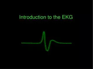
INTRODUCTION. 12-Lead EKG machine developed in 1903 timeless invention Inexpensive, easily accessibleGoals1. Review basic cardiac physiology2. Develop systematic approach to 12-Lead interpretation3. Practice interpreting EKG strips . 12-Lead EKG. Electrical recording of the heart's electrical activityCardiac cells

E N D
1. Introduction to the basics of 12-Lead EKG Interpretation Jennifer Rodgers, MSN, ARNP
Wichita State University
Summer 2006
2. INTRODUCTION 12-Lead EKG machine developed in 1903 timeless invention
Inexpensive, easily accessible
Goals
1. Review basic cardiac physiology
2. Develop systematic approach to 12-Lead
interpretation
3. Practice interpreting EKG strips
Remarkable clinical power
One glance>diagnose evolving MI, arrhythmia, re-assurance for patient wanting to start exercise program, OR chronic effects of HTN
Only a tool>reliable as userRemarkable clinical power
One glance>diagnose evolving MI, arrhythmia, re-assurance for patient wanting to start exercise program, OR chronic effects of HTN
Only a tool>reliable as user
3. 12-Lead EKG Electrical recording of the heart�s electrical activity
Cardiac cells �resting state polarized (negative inside positive outside)
Ensure appropriate distribution of ions (potassium, sodium, chloride, calcium)
Depolarization-fundamental electrical event of the heart, stimulation>muscle begins to work
Propagated from cell to cell>wave throughout entire heart>flow of electricity Currents can be detected by external electrodes
All the various waves seen on EKG>manifestations of depolarization and polarizationCurrents can be detected by external electrodes
All the various waves seen on EKG>manifestations of depolarization and polarization
4. TYPES OF CELLS PACEMAKER-electrical source
ELECTRICAL CONDUCTING-hard wiring
MYOCARDIAL-contractile machinery
5. PACEMAKER CELLS Small 5-10 cm in length
Depolarize spontaneously @ particular rate
Located in Right Atrium-Sinoatrial (sinus) node
Typical 60-100 beats/minute
Dependent on autonomic nervous system and body demands
Anything that affects cardiac output
Cycle polarization-depolarization>action potentialAnything that affects cardiac output
Cycle polarization-depolarization>action potential
6. ELECTRICAL CONDUCTING CELLS
Long-Thin cells
Rapidly carry currents to distant regions of heart
7. Myocardial Cells Heavy labor cells
Constantly contracting & relaxing> delivering blood to the periphery
Contain contractile proteins> actin & myosin
Depolarization>myocardial cell>calcium released within cell>contract
8. Time & Voltage Waves on EKG primarily reflect electrical activity>myocardial cells
Waves-3 characteristics
1. Duration-measured fraction/second
2. Amplitude-measured millivolts (mV)
3. Configuration-shape/appearance
9. EKG PAPER Light lines small squares- 1 X 1 mm
Bold lines large squares 5 X 5 mm
Horizontal axis=time
1. Distance across small square=0.04 sec.
2. Distance across large square=0.2 sec.
Vertical axis=voltage
1. Distance across small square=0.1 mV
2. Distance across large square=0.5 mV
6 second strip to figure rate (X 10) (30 lg=6)
10. SINUS NODE STARTS EACH CARDIAC CYCLE OF CONTRACTION & RELAXATION BY SPONTANEOUS DEPOLARIZATION THIS IS NOT SEEN ON THE EKG
SA NODE FIRES>DEPOLARIZATION BEGINS>ATRIAL CONTRACTION (LIKE PEBBLE IN POND)
ELECTRODES MEASURES ATRIAL POLARIZATION/DEPOLARIZATION> P WAVESA NODE FIRES>DEPOLARIZATION BEGINS>ATRIAL CONTRACTION (LIKE PEBBLE IN POND)
ELECTRODES MEASURES ATRIAL POLARIZATION/DEPOLARIZATION> P WAVE
11. ATRIOVENTRICULAR (AV) NODE Electrical Gatekeeper between atria and ventricles
Allows atrial contraction to end & empty contents into the ventricle before ventricular contraction begins ELECTRICAL GATEKEEPER JUNCTION ATRIA & VENTRICLES
AV NODE> SLOWS CONDUCTION TO A HALT> ALLOWS ATRIAL CONTRACTION END & EMPTY CONTENTS INTO VENTRICULE BEFORE VENTRICULAR CONTRACTION BEGINSELECTRICAL GATEKEEPER JUNCTION ATRIA & VENTRICLES
AV NODE> SLOWS CONDUCTION TO A HALT> ALLOWS ATRIAL CONTRACTION END & EMPTY CONTENTS INTO VENTRICULE BEFORE VENTRICULAR CONTRACTION BEGINS
12. VENTRICULAR DEPOLARIZATION WAVE DEPOLARIZATION SPREADS THROUGH THE 3 PARTS-Bundle of His (intrinsic 40-60 bpm)> Bundle Branches> Purkinje Fibers (intrinsic 20-40 bpm) & out into the ventricular myocardium
Beginning ventricular depolarization>QRS complex Amplitude QRS much greater than P wave>ventricles are much larger
First deflection Q WAVE
First upward deflection R WAVE
Next downward deflection S WAVE
Amplitude QRS much greater than P wave>ventricles are much larger
First deflection Q WAVE
First upward deflection R WAVE
Next downward deflection S WAVE
13. VENTRICULAR REPOLARIZATION Brief refractory period
Restore electro negativity of their interiors
�T wave�
Atrial repolarization is not seen
14. PR INTERVAL Includes P wave & the first straight line connecting it to the QRS interval
Measures the time from the start of atrial depolarization to the start of ventricular depolarization
Normal 0.12-0.20 sec
>0.20 delay in AV conduction
<0.12 shortens as HR increases
15. ST SEGMENT The straight line connecting the end of the QRS complex with the beginning of the T wave
Measures the time from the end of ventricular depolarization to the start of ventricular depolarization
16. QT INTERVAL Includes the QRS complex, ST segment, & T wave
Measures the time from the beginning of ventricular depolarization to the end of ventricular repolarization
Normal duration QRS 0.06-0.10 seconds
17. RATE MEASURMENT 1. COUNT THE # OF QRS COMPLEXES IN 6 SECONDS X 10, MOST COMMON
2. COUNT # OF LG. BOXES BETWEEN 2 R WAVES /BY 300
3. COUNT # OF SM. BOXES BETWEEN 2 R WAVES /BY 150
1. EASIEST CAN BE USED WITH ANY TYPE RHYTHM , SIMPLE, QUICKEST1. EASIEST CAN BE USED WITH ANY TYPE RHYTHM , SIMPLE, QUICKEST
18. STEPWISE APPROACH STRIP INTERPRETATION A. Determine Atrial & Ventricle Rate
1. V-measure R-R, A-measure P-P
2. >100 Tachycardia, <60 Bradycardia
B. R-R Interval Regular?
C. P wave Formation
1. Precede QRS, occur regularly,
similar size
2. P wave + (SA Node) OR -/absent (AV
Junction)
D. QRS wide or narrow
19. SINUS NODE DYFUNCTION SINUS ARRHYTHMIA
SINUS TACHYCARDIA
SINUS BRADYCARDIA
20. SINUS ARRHYTHMIA A. Rate 60-100 bpm
B. *R-R irregular
C. *Normal P wave
D. Normal PR interval 0.12-0.20 sec.
E. Normal QRS complex </=0.10 sec.
Phasic slowing & quickening, benign, normal response to respirations, asymptomatic
Except in elderly>Sick Sinus Syndrome, not usually seen in infants INSPIRATION>INCREASE BLOOD FLOW>HR INCREASE, EXPIRATION>DECREASED VENOUS RETURN>HR SLOWSINSPIRATION>INCREASE BLOOD FLOW>HR INCREASE, EXPIRATION>DECREASED VENOUS RETURN>HR SLOWS
21. SINUS BRADYCARDIA Usual response to reduced demand for blood flow
A. *Rate < 60 bpm
B. R-R Regular
C. Normal P wave
D. Normal PR interval
E. Normal QRS Complex
Asymptomatic Vs. Symptomatic EXAMPLE: NORMAL ATHLETES>CONDITIONED, AMI INFERIOR >FAVORABLE UNLESS HYPOTENSION , OFTEN MEDICATION INDUCED >BBEXAMPLE: NORMAL ATHLETES>CONDITIONED, AMI INFERIOR >FAVORABLE UNLESS HYPOTENSION , OFTEN MEDICATION INDUCED >BB
22. SINUS TACHYCARDIA ACCELERATION SA NODE
A. *Rate >110 bpm (110-160)
B. R-R Regular
C. Normal P wave
D. Normal PR Interval 0.12-0.20 sec
E. Normal QRS Complex </=0.10 sec
Response to exercise/stress, OR response illness (hypovolemia/hypotension)>resolves once cause fixed
EXAMPLE 67 FEMALE (TURNER) LUNG CA> ST 150�S, HYPOXEMIA, BP 80/40, ABG>ACUTE RESP. FAILURE, CXR>RML INFILTRATE + BACTEREMIA >BIPAP>30 MINUTES HR 110
EXAMPLE 67 FEMALE (TURNER) LUNG CA> ST 150�S, HYPOXEMIA, BP 80/40, ABG>ACUTE RESP. FAILURE, CXR>RML INFILTRATE + BACTEREMIA >BIPAP>30 MINUTES HR 110
EXAMPLE 67 FEMALE (TURNER) LUNG CA> ST 150�S, HYPOXEMIA, BP 80/40, ABG>ACUTE RESP. FAILURE, CXR>RML INFILTRATE + BACTEREMIA >BIPAP>30 MINUTES HR 110
EXAMPLE 67 FEMALE (TURNER) LUNG CA> ST 150�S, HYPOXEMIA, BP 80/40, ABG>ACUTE RESP. FAILURE, CXR>RML INFILTRATE + BACTEREMIA >BIPAP>30 MINUTES HR 110
23. ATRIAL DYSRHYTHMIAS Most common cardiac rhythm disturbance
Originate in/around SA Node �above ventricle�
Can diminish atrial �kick� >20% ventricular volume
PSVT-Paroxysmal Supraventricular Tachycardia
Atrial Fibrillation
Atrial Flutter
24. PSVT A. Rate 150-250 bpm
B. *Regular R-R interval
C. P wave can be buried
D. PR interval may be hard to find
E. *Normal �narrow� QRS complex
Treatment LVEF50%>CCB, BB, Dig., possible cardioversion, <40% No Cardioversion!, Dig., Amiodorone, Diltiazem
25. ATRIAL FLUTTER A. Atrial Rate 250-350 bpm
B. R-R Irregular
C. *P wave �classic saw tooth OR flutter�
D. PR interval immeasurable
E. QRS complex narrow
May have palpitations, OR s/sx reduced C.O.
If symptomatic>cardioversion, BB, Sotalol, Dig.
26. ATRIAL FIBRILLATION Chaotic, asynchronous electrical activity in atrial tissue>multiple impulses numerous eptopic pacemakers
A. Atrial Rate-indiscernible, V-Rate 60-160 (RVR-Rapid Ventricular Response)
B. *R-R Irregular
C. *No P wave
D. No PR interval
E. QRS narrow PREVELANCE 8-10% >75, RESPONSIBLE 15-20% CVA�S, LEADING CAUSE CVA ELDERLY
KNOW TREATMENT, DON�T FORGET ANTICOAGULATIONPREVELANCE 8-10% >75, RESPONSIBLE 15-20% CVA�S, LEADING CAUSE CVA ELDERLY
KNOW TREATMENT, DON�T FORGET ANTICOAGULATION
27. JUNCTIONAL ESCAPE RHYTHM Originates in AV junction �escape pacemaker�
A. Rate 40-60 bpm
B. R-R Regular
C. *Inverted P wave, preceding each QRS
D. *PR Interval short 0.10 sec.
E. QRS normal
How is patient tolerating? Loss of �atrial kick�> can reduce C.O. by 20%
28. PREMATURE BEATS Premature Atrial Contractions (PAC�s)-originate outside AV node, single/multiple ectopic focus supersede SA node
Premature Ventricular Contractions (PVC�s)-ectopic beats that originate in ventricles & occur earlier, singles, pairs or in clusters
29. VENTRICULAR DYSRHYTHMIAS
VENTRICULAR TACHYCARDIA
VENTRICULAR FIBRILLATION
30. VENTRICULAR TACHYCARDIA (V-TACH) Defined as: Vent. Rate > 100 bpm & when 3 OR more PVC�s strike in a row
Life threatening, unstable, sustained OR unsustained
A. A-rate can�t be determined, V-rate100-250 bpm
B. R-R regular or slightly irregular
C. P wave usually absent, dissociated
D. PR Interval-immeasurable
E. *WIDE QRS >0.12 sec. Bizarre appearance Don�t want to miss unpredictableDon�t want to miss unpredictable
31. VENTRICULAR FIBRILLATION (V-FIB) VF-Full cardiac arrest, no pulse/BP, always check patient first>Defibrillate>CPR/ACLS
A. Rate-can�t be determined pulseless
B. R-R can�t determine
C. P wave can�t be determined
D. PR interval can�t be determined
E. QRS complex can�t be determined
Ventricular electrical activity >fibrillatory waves with no recognizable pattern
32. A-V BLOCKS Interruption/delay in the conduction of electrical impulses between the atria & ventricles
Classified site of block/severity of conduction abnormality
1st degree, 2nd degree Mobitz I (Wenkebach), 2nd degree Mobitz II, 3rd degree (Complete heart block)
33. 1st Degree AV Block Characterized by PR Interval > 0.20 seconds
Delay in conduction AV Node
Prolonged PR Interval constant
Usually asymptomatic
Least concerning of the blocks
34. 2nd Degree Mobitz I (Wenkebach) Successive impulses from SA node delayed slightly longer than the previous impulse
Characterized by prolonged PR interval that continues until the P wave is dropped (impulse doesn�t reach ventricle)
May have hypotension or lightheadedness
35. 2nd Degree Mobitz II Less common, more serious
Impulses from SA node fail to conduct to ventricles
Hallmark PR Interval constant normal or prolonged, doesn�t prolong before dropping, not followed by QRS, can have > 1 dropped in a row
Precursor to 3rd Degree Heart Block P-P CONSTANT EVEN IN DROPPED BEATP-P CONSTANT EVEN IN DROPPED BEAT
36. 3RD DEGREE �COMPLETE HEART BLOCK� Indicates complete absence of impulse between the atria & ventricle
Atrial rate > or = ventricular rate
Occur @ AV node 40-60 bpm
Occur @ bundle branches < 40 bpm wide QRS complex
Decreased C.O., P-P & R-R disassociated
37. EKG INTERPRETATION
LET�S PRACTICE!!!!