Download
1 / 53
530 likes | 542 Vues
Prof. Amgad Fouad Gastroenterology center Mansoura University. Achalasia of the esophagus, different line for therapy. Achalasia Greek word Failure to relax Willis (1672). Zenker & Vonziemssen (1877): Diminished contractile power of the esophageal musculature.

E N D
Prof. Amgad Fouad Gastroenterology centerMansoura University
Achalasia • Greek word • Failure to relax • Willis (1672)
Zenker & Vonziemssen (1877):Diminished contractile power of the esophageal musculature. • Meltzer & Miklicz (1888):Spasmodic contraction of the cardiac sphincter. • Einhorn (1888):Failure of relaxation of the cardia on swallowing. • Horst (1929):Established the term achalasia (failure to relax).
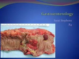
Achalasia: • The most recognised motor disorder of the esophagus. • Cardinal features: • Poorly relaxing LES • Prolonged esophageal transit • Defective esophageal body peristalsis .
1-2 / 200.000 • ♂ =♀ . • Any age. • Onset 3rd – 5th decade. • Duration of symptoms at presentation 2 years average. (Mayberry & Atkinson, 1985)
Presentation • Dysphagia(almost 100%) • Regurgitation (60-90%) • Chest pain(30 – 50%) • Wt loss (Advanced disease). • Pulmonary symptoms. • Bronchopneumonia • Lung abscess • Ht burn (rare presentation )
Diagnosis • Compatible clinical history. • Radiography. • Endoscopy. • Manometry.
Radiographic studies Plain X ray : • Widened mediastinum . • Air fluid level • Absence of gastric air bubble • Evidence of pulmonary complications
Radiographic studies(continue) Barium swallow • Screening test • Esophageal body dilated • Lower end esophagus → tapered point. (birds Beak ) • Nitrite test → Diagnostic.
Radiographic staging • Stage I :Slight dilatation of the body not >3.5cm. • Stage II :Moderate dilatation 3.5-6cm. • Stage III :Marked dilatation >6cm. • Stage IV :Marked dilatation elongation and tortiousity of the esophagus (sigmoid esophagus).
Endoscopy Important diagnostic tool • Rule out several diseases that mimic achalasia. • Evaluate esophageal mucasa before therapeutic manipulation. Typical finding • Dilated esophgeal body • Puckered closed LES • No organic stricture
Mamometry • Confirms & establishes the diagnosis • Features: Essential features • Absence of esophgeal body peristalsis (1ry peristaltic waves) • ↑ intraesophageal resting pressure. • Abnormal LES relaxation. Supportive Features • Hypertensive LES pressure • Low amplitude esophageal contractions (Castell, 1996)
Medical Treatment Nitrates . Ca ++ channel blockers Anticholinergic drugs • All have been shown to reduce the force of contraction of esophageal body smooth muscles. • May be of value in reducing chest pain & improving dysphagia (Gelfond et al ., 1982)
Botulinum toxin • Neurotoxin produced by clostridium botulinum. • Only serotype A+B have been approved for clinical use. • Two preparations of BTX (A) available: • Dysport (UK) • Botox (USA)
Pneumatic dilatation • The most effective non-surgical treatment option. • Forceful dilatation using air or water pressure can be applied to the lower esophageal segment and controlled to secure further stretching to the point of rupture of the circular ms fibers.
Recommended technique for pneumatic balloon dilatation (Voizi et al, 1994)
Cumulative effectiveness of pneumatic dilatation in Achalasia
Surgical management of Achalasia • Rationale of surgery is to weaken the lower esophageal sphincteric pressure but in controlled Faison avoiding subsequent reflux (Earlam, 1976)
Heller (1913) • First performed extra mucosal cardiorytomy. • Two 8cm incisions one ant & one post. • Incisions extending 2cm into the dilated part cranially & into the fundus of the stomach caudally.
Modified Heller’s myotomy (Zaaijer, 1985) • Most widely used technique. • Single anterior myotomy. • Transabdominal. However Two problems →poor results • Incomplete myotomy • Reflux esophagitis .
Laparoscopic Heller’s • Most preferred by gastrointestinal laparoscopic surgeon. • Easy access. • Good result • Anti – reflux +
Aim of work • The aim of this work was to evaluate Heller’s myotomy and preumatic balloon dilatation as two alternative lines of therapy for patients with achalasia of the esophagus.
Our study is a retrospective non- randomised study conducted at GEC during the period between October 1979- November 2002. • The study included 310 cases with achalasia. • 169 ♂ & 141♀ • According to the line of management the study included two groups: • Group A: 150 patients treated with myotomy + fundoplication. • Group B: 160 patient treated by pneumatic balloon dilatation.
Preoperative work up • Thorough history and clinical examination. • Patients were divided into 4 groups according to Demeester's grading for dysphagia. (Cuschieri et al., 2002) • No dysphasia • Mild : occasional episodes. • Moderate: requires fluids to clear. • Severe : episodes of solid food impaction & require medical treatment.
Preoperative work up(continue ) • Radiological examination • According to Olsen scoring system, patients were divided into 4 groups (Olsen et al., 1983) • Endoscopic evaluation • Manometric study
Method of management Group (A): • Modified Heller myotomy35 patient (56.8%). • Myotomy + Dor fundoplication45 patient (30%). • Myotomy + Nissen fundoplication9 patient (6%). • Myotomy + Taupet fundoplication4patient (2.6%). • Laparoscopic mytomy3 patients (2%). • Laparoscopic mytomy + Dor fundoplication 4patints (2.6%).
Method of management Continue Group (B): • Pneumatic balloon dilatation (1.8+ 1 set) • One session in 79 patients (60.5%). • Two sessions in 48 patients (30%). • Three or more sessions in only 15 patients (9.5%).
Radiological characters of achalasia patients before management .
Endoscopic findings in patients with achalasia before management .
Symptomatic evaluation of patients after pneumatic ballon dilatation .