Download
1 / 50
500 likes | 1.24k Vues
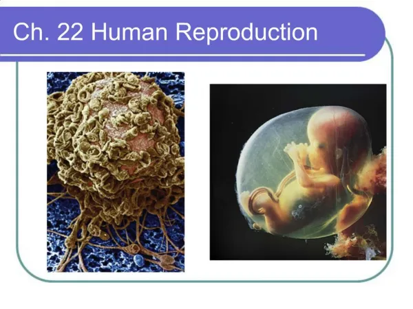
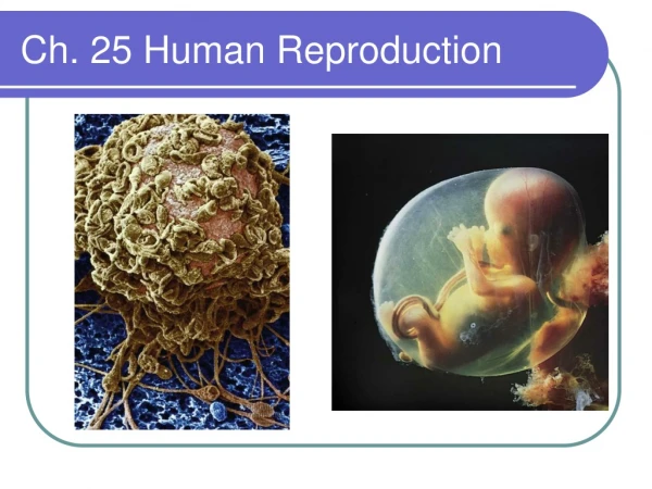
Ch. 25 Human Reproduction. Section Objectives. Identify the structures and functions of the male and female reproductive systems. Summarize the internal feedback control of reproductive hormones Sequence the stages of the menstrual cycle. main functions

E N D
Section Objectives • Identify the structures and functions of the male and female reproductive systems. • Summarize the internal feedback control of reproductive hormones • Sequence the stages of the menstrual cycle.
main functions 1. the production of sperm—the male sex cells 2. their delivery to the female. Reproductive anatomy of the human male
Reproductive anatomy of the human male • Semen • Sperm, which are expelled through the ducts during ejaculation • Glandular secretions that carry, nourish, and protect the sperm • Testes • Produce sperm • Located outside abdominal cavity within the scrotum (saclike pouch~ 1-3°C below normal body temperature- sperm can only form at this lower temp.)
How sperm leave the testes • . Seminiferous tubules: carries/stores sperm in testes • 2.Epididymis: a series of coiled ducts for maturation & temporary storage organ for sperm • 3. Vas deferens: tube which carries sperm past lubricating glands • 4. ( a.)Seminal vesicles: secrete fluid that protects & nourishes sperm (b.) Prostrate gland: produces an alkaline fluid that neutralizes urine in the urethra (c.) Bulbourethral glands: secrete fluid that may help lubricate the urethra 5. Urethra: tube in the penis that transports sperm out of the male’s body, also transports urine from the urinary bladder. 6. Penis: copulatory organ, releases semen 7. Ejaculation: the release of semen
main functions of the female reproductive system 1. to produce eggs, the (female sex cells), 2.to receive sperm, 3. to provide an environment in which a fertilized egg can develop. Reproductive anatomy of the human female
Ovaries Contain follicles that nurture eggs (ova) Produce sex hormones Functional from puberty to menopause Oviducts (Fallopian Tubes) Convey eggs to the uterus Muscular contractions &cilia draw ovum (egg) into oviduct Fertilization occurs Reproductive anatomy of the human female
Uterus (womb) Development of fertilized egg Opens into the vagina Vagina Receives penis during intercourse Forms the birth canal Reproductive anatomy of the human female
Puberty • Puberty: when secondary characteristics develop and the potential for sexual reproduction is reached(sperm production or ovulation) • Changes are controlled by hormones secondary sex characteristics: Males: hormone~ testosterone, characteristics~ body hair, muscle development, deep voice Females: Hormone~ estrogen. Characteristics~ breasts, broadened pelvis, distribution of body fat
Reproductive hormones • Testosterone • from testes • sperm production & secondary sexual characteristics • Estrogen • from ovaries • egg production, preparing uterus for fertilized egg & secondary sexual characteristics
Androgens (testosterone most important) stimulate sperm production They also maintain homeostasis by a negative feedback mechanism that inhibits the secretion of FSH (follicle-stimulating hormone) and LH (luteinizing hormone) Hormonal control of the testes Stimuli from otherareas in the brain Hypothalamus Releasinghormone Anteriorpituitary Negative feedback FSH LH Androgenproduction Testis Spermproduction
Oogenesis: Production of eggs • Most of the process occurs within the ovaries • Lifetime supply of primaryoocytes is present at birth • One primary oocyte matures each month to form a secondary oocyte • If the secondary oocyte is fertilized, it completes meiosis and becomes a haploid ovum
Egg maturation in ovary releasesprogesterone maintainsuteruslining produces estrogen
Menstrual Cycle • The series of changes in the female reproductive system that includes producing an egg and preparing the uterus for receiving it. • Once an egg has been released during ovulation, the part of the follicle that remains in the ovary develops into a structure called the corpus luteum. • The menstrual cycle begins during puberty and continues for 30 to 40 years, until menopause. • At menopause, the female stops releasing eggs and the secretion of female hormones decreases.
The Reproductive Cycle of the Human Female • A cyclic pattern of hormone secretion and reproductive events. • Humans and many other primates have menstrual cycles. • If pregnancy does not occur the endometrium (lining of uterus) is shed through the cervix and vagina: menstruation
divided into three phases: the flow phase, the follicular phase, and the luteal phase. The timing of each phase of the menstrual cycle correlates with hormone output from the pituitary gland, changes in the ovary, and changes in the uterus The Menstrual Cycle
Menstrual cycle LH • Controlled by interaction of 4 hormones • FSH & LH • estrogen • progesterone FSH ovulation = egg release egg development corpus luteum estrogen progesterone lining of uterus days 0 7 14 21 28
Flow Phase • Day 1 of the menstrual cycle is the day menstrual flow begins • the shedding of blood, tissue fluid, mucus, and epithelial cells that made up the lining of the uterus, the endometrium. • Contractions of the uterine muscle help expel the uterine lining and can cause discomfort in some females. • the level of FSH in the blood begins to rise, and a follicle in one of the ovaries begins to mature as meiosis of the prophase I cell proceeds.
Follicular Phase: second phase of the menstrual cycle • lasts from about day 6 to day 14. • As the follicle containing a primary oocyte continues to develop, it secretes estrogen, which stimulates the repair of the endometrial lining of the uterus. • Day 14 ovulation occurs~ follicle enlarges and ruptures ovary wall. Egg is released to oviduct.
Luteal Phase • Progesterone increases the blood supply of the endometrium • These changes correspond to the arrival of a fertilized egg. • If the egg is not fertilized, the rising levels of progesterone and estrogen from the corpus luteum cause the hypothalamus to inhibit the release of FSH and LH. • The corpus luteum degenerates and stops secreting progesterone or estrogen. • As hormone levels drop, the thick lining of the uterus begins to shed. • If fertilization occurs the endometrium begins secreting a fluid rich in nutrients for the embryo.
corpusluteum ovary yes corpusluteum no Female reproductive cycle Feedback eggmatures & is released(ovulation) builds up uterus lining estrogen progesterone FSH & LH fertilized egg(zygote) maintainsuterus lining HCG pituitarygland pregnancy progesterone GnRH corpus luteum breaks down progesterone drops menstruation maintainsuterus lining hypothalamus
Female hormones • FSH & LH • released from pituitary • stimulates egg development & hormone release • peak release = release of egg (ovulation) • Estrogen • released from ovary cells around developing egg • stimulates growth of lining of uterus • decreasing levels causes menstruation • Progesterone • released from “corpus luteum” in ovaries • cells that used to take care of developing egg • stimulates blood supply to lining of uterus • decreasing levels causes menstruation
FSH (follicle stimulating hormone) produced by pituitary stimulates development of follicle LH (luteinizing hormone) stimulates the development of the corpus luteum, stimulates ovulation Estrogen: secreted by ovaries, stimulates development of uterine lining Progesterone: secreted by corpus luteum, maintains uterine lining Hormonal coordination of the menstrual and ovarian cycles .
Ch. 25 • Section Objectives • Describe the processes of fertilization and implantation. • Summarize the events during each trimester of pregnancy.
Fertilization results in a zygote and triggers embryonic development • Fertilization is the union of a sperm and an egg to form a diploid zygote (PATH) Millions of sperm ->vagina -> cervix -> uterus -> oviduct (site of fertilization) sperm + egg -> zygote 23(n) + 23(n) -> 46(2n)
Fertilization • Only one of these sperm will penetrate this human egg cell to initiate fertilization • The shape of a human sperm cell is adapted to its function
Implantation • Implantation: fertilized egg implants in thickened uterine lining the embryo starts to secrete the hormone human chorionic gonadotropin (HCG) (hormone for pregnancy tests) This hormone keeps the corpus luteum alive so that it continues to secrete progesterone • By the third or fourth month, the placenta takes over for the corpus luteum, secreting enough estrogen and progesterone to maintain the pregnancy.
Embryonic Development • Development: series of orderly, precise steps that transform a zygote into a multicellular embryo ~early stage of development of multicellular organism • Includes: 1. cell division 2. cell growth 3. cell differentiation ~changing of unspecialized embryonic cells into specialized cells, tissues,& organs
Early Embryonic Development • Cleavage is the first major phase of embryonic development • It is the rapid succession of cell divisions (Mitotic) • It creates a multicellular embryo from the zygote • NO growth • Stages: • 1. Morula~solid ball of cells • 2.Blastula~ single layer of cells surrounding a fluid-filled cavity called the blastocoel ZYGOTE Blastocoel Cross sectionof blastula BLASTULA(hollow ball)
Embryonic Development • Gastrulation is the second major phase of embryonic development • The cells at one end of the blastula move inward • Organs start to form after gastrulation • Embryonic tissue layers begin to differentiate into specific tissues and organ systems
Embryonic Membranes and the Placenta • Amnion~fluid filled sac for protection • Chorion ~ will form the embryo’s part of the placenta • Yolk sac ~ produces first blood cells & germ cells • Allantois ~ will form the umbilical cord (ropelike structure that attaches embryo to uterus) • Placenta~A growing fetus exchanges nutrients, oxygen, and wastes with the mother through the placenta. Chorion Amnion Allantois Yolk sac
Placenta • Food & gases diffuse across blood vessels
Fetal Development • Gestationis pregnancy • It begins at conception and continues until birth • Pregnancy in humans usually lasts about 280 days, calculated from the first day of the mother’s last menstrual period. • Embryonic development of essential organs occur in early pregnancy • The embryo may encounter risks from faults in its genes & from mother’s exposure to environmental factors
Human development from conception to birth is divided into three trimesters • First trimester • First three months • The most rapid changes occur during the first trimester 10 weeks • 10 weeks10 weeks • 10 weeks 10 weeks 4 weeks 7 weeks
Human development from conception to birth is divided into three trimesters • Second trimester • Increase in size of fetus • General refinement of human features 12 weeks
Human fetal development • The fetus just spends much of the 2nd & 3rd trimesters just growing …and doing various flip-turns & kicks inside amniotic fluid Week 20
Human fetal development • 24 weeks (6 months; 2nd trimester) fetus is covered with fine, downy hair called lanugo. Its skin is protected by a waxy material called vernix
Human fetal development • 30 weeks (7.5 months) umbilical cord
Getting crowded in there!! • 32 weeks (8 months) The fetus sleeps 90-95% of the day & sometimes experiences REM sleep, an indication of dreaming
Human development from conception to birth is divided into three trimesters • Third trimester • Growth and preparation for birth
Section Objective: • Describe the three stages of birth. • physiological and physical changes a female goes through to give birth are called labor. • Labor begins with a series of contractions of the uterine muscles. • These contractions are stimulated by oxytocin, a hormone released by the pituitary.
Birth positive feedback
Three stages of labor • .Dilation of the cervix is the first stage -Cervix reaches full dilation at 10cm • Longest stage of labor (6-12 hours or longer)
Three stages of labor • Expulsion is the second stage • Period from full dilation of the cervix to delivery of the infant • Uterine contractions occur every 2-3 minutes • Mother feels urge to push down with her abdominal muscles • Infant is forced down and out of uterus and vagina within a period of 20 minutes
Three stages of labor • The delivery of theplacenta is the final stage of labor • Usually occurs within 15 minutes after the birth of the baby
The end of the journey! And you think 9 months of Biology is hard!
Growth and Aging • Once a baby is born, growth continues and learning begins • Human growth varies with age and is somewhat gender dependent.
An adult ages • As an adult ages, his or her body undergoes many distinct changes. • Slower metabolism • White hair • Thinner bones • Vision & hearing dimminish