Pathologic Fractures – Metastasis
Pathologic Fractures – Metastasis. Orthopedic Surgery Grand Round 7 th February 2013 Dr. J.W. Kinyanjui Registrar Ward 6D. Outline. Introduction Epidemiology Pathophysiology Clinical evaluation Management. Introduction. Fracture through abnormal bone

Pathologic Fractures – Metastasis
E N D
Presentation Transcript
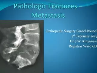
Pathologic Fractures – Metastasis Orthopedic Surgery Grand Round 7th February 2013 Dr. J.W. Kinyanjui Registrar Ward 6D
Outline • Introduction • Epidemiology • Pathophysiology • Clinical evaluation • Management
Introduction • Fracture through abnormal bone • Minor trauma or during normal activity • 5th decade most prevalent • Metastases 2nd most common cause of pathologic fractures • F: breast and lungs – 80% • M: prostate and lungs – 80% • 10% - no primary tumor found
Epidemiology – incidence at autopsy Primary Site% metastasis to Bone Breast 50-85 Lung 30-50 Prostate 50-70 Hodgkin’s 50-70 Kidney 30-50 Thyroid 40 Melanoma 30-40 Bladder 12-25
Pathophysiology • Most spread is hematogenous • Few tumors due to contiguous spread • Most common osteolytic via osteoclast stimulation • Prostate – commonly osteoblastic • Breast – mixed • Theories explaining predilection of bone for metastasis
Paget’s fertile soil hypothesis • 1889 • Sites of secondary growths are not a matter of chance • Some organs provide a more fertile environment for the growth of certain metastases • Example: breast cancer to liver, Krukenberg tumor • Prostate cancer to bone • Hart and fielder later proved this using radioactive labelling
Ewing’s circulation theory • 1928 • Metastatic deposits dependent on route of blood and lymph flow • Organs though to be passive receptacles • Organs with prominent venous systems have more secondaries • Baston plexus of spine responsible for prostate secondaries
Red marrow theory • In descending order of frequency: • Spine • Pelvis • Ribs • Proximal appendicular skeleton • Marrow sinusoids more susceptible to tumor cell penetration • Sudden change from arterioles to sinusoids favours tumor cell entrapment • Ewing’s and Paget’s theories not mutually exclusive
Molecular level • Cells from primary enter blood vessels • Attachment and penetration of basement membrane, neovascularisation • Type 1 collagen shown to be chemotactic to tumor cells • RANK ligand produced by tumor cells stimulating osteoclast activity • PTHrP produced by breast and lung cancer cells stimulates osteoclasts • Prostate cancer cells produce BMPs, IGF1, TGFβ2 which stimulate osteoblasts
Clinical evaluation: History • Pain – most common, preceding fracture, night, constant, dull, aggravated by activity • Trauma – usually minimal for type of fracture • Constitutional – anorexia, night sweats, weight loss, fatigue • Previous cancer • Carcinogen – smoking, radiation, occupational toxins
Factors suggesting pathologic fracture • Spontaneous fracture • Minor trauma • Pain at site preceeding fracture • Multiple recent fractures • Age > 45 yrs • Prior history of malignancy
Associated problems • Lowered Quality of life: • Debilitating pain • Immobility • Neurologic deficits – spine mets • Anaemia • Hypercalcemia
Hypercalcemia • Neurologic: headache, confusion, irritability, blurred vision • Gastrointestinal: anorexia, nausea, vomiting, abdominal pain, constipation, weight loss • Musculoskeletal: fatigue, weakness, joint and bone pain, unsteady gait • Urinary:nocturia, polydypsia, polyuria, urinary tract infections
Clinical evaluation: examination • Local: mass, deformity, tenderness, contiguous skeleton, neurologic exam • Systemic: cachexia, pallor, lymphadenopathy, entire skeletal system • Primary: breast, thyroid, prostate, lung, pelvic
Clinical evaluation: Laboratory • TBC – anaemia of chronic disease • Calcium – elevated • Alkaline phosphatase – elevated, non specific • Tumor markers – PSA, CEA, CA125, TFTs • N-telopeptide + C-telopeptide – markers of bone destruction, determine extent of skeletal involvement, assess response to bisphosphonates
Imaging: plain radiographs • Enneking’s questions: • Location: diaphysis, metaphysis, epiphysis, cortical or medullary • Effect: osteoblastic vs. osteolytic or mixed • Reaction: sclerotic rim, periosteal reaction, codman triangle • Isolated avulsion of lesser trochanter – imminent femoral neck fracture
Radiology: CT scans • Most sensitive for detecting bone destruction • Determines extent of cortical involvement • Also used to search for primary lesion in pelvis, abdomen or chest
Radiology: MRI • Most sensitive for assessment of the anatomic extent of a lesion • Most adequate for spinal metastases to determine neurologic structure involvement • Can determine extraosseous spread of a mass
Bone scanning • Technetium-99m (99m Tc) bone scanning: • Sensitive for detection of occult lesions • Assessment of the biologic activity of lesions • Identification of other sites • Assessing response to therapy
Biopsy • Indicated to rule out primary tumor of bone • Immunohistochemistry can determine primary • Biopsy at fracture site complicated by bleeding and callus formation • Needle vsincisional • Oncological surgical principles adhered to • Cultures to rule out infection
Impending pathologic fractures • Prophylactic stabilisation before radiotherapy can be performed for pain • Radio and chemotherapy without stabilisation also an option • Decision to stabilise difficult • Mirel’s criteria useful to determine which lesions at high risk of fracture
Mirel’s criteria Size is the diameter of cortex involved on plain radiographs A score of 8 or more is an indication for prophylactic stabilisation
Advantages • Prophylactic stabilisation: • Shorter hospital stay • More immediate pain relief • Faster and less complex surgery • Quicker return to premorbid function • Improved survival
Management objectives • Decrease pain • Restore function • Maintain/restore mobility • Limit surgical procedures • Minimize hospital time • Early return to function (immediate weightbearing)
Non operative management • Bisphosphonates – modifies bone resorption by osteoclasts, shown to reduce risk of skeletal metastasis • Hematologic – correction of anaemia, coagulopathy, DVT prophylaxis • Hypercalcemia – hydration, calcium restriction, bisphosphonates, mithramycin • Analgesia • Radiation – most useful in spinal metastases
Radiotherapy • Used to reduce pain secondary to bone metastases • Partial in 80%. Complete in 50 – 60% • Halts progression of bony destruction • Allows healing of an impending pathologic fracture • Postoperative local tumor control
Bracing • Patients with limited life expectancies, severe comorbidities, small lesions, or radiosensitive tumors • Upper extremity lesions particularly amenable • Adjuvant radiotherapy of suscepible tumors required
Operative: principles • Durable, weight bearing impalnts needed • PPMA augmentation of construct useful incl. prosthesis • Bone graft less useful due to prolonged healing time • Prophylacticallystabilise as much bone as possible • Anticipate hemorrhage due to neovascularisation • Thus tourniquet, preoperative embolisation
Upper extremity • Scapula, clavicle – non operative • Proximal humerus – prosthesis (long stem), intramedullary nail with multiple screws • HumerusDiaphysis – locked IM nail > plating • Distal humerus – prosthesis, retrograde flexible IM nails > bicondylar plating • Forearm – Rare. IM nails or plating
Lower extremity • Acetabular – reconstruction with appropriate prosthesis • Femoral neck – hemi- or THR. Cemented. Long stem • Intertrochanteric – recon nail or prosthesis > DHS • Subtrochanteric – locked IM nail • Femur shaft – locked IM nail preferably cephalomedullary • Around the knee – locked plating > retrograde nailing
Spinal fractures • Commonly present with compression fracture • MRI to differentiate from osteoporosis • Lesion involving body and pedicle sparing disc highly suggestive • Radiotherapy, steroids if no neurodeficits or impending fracture
Spinal fractures • Surgery: • Progression of disease after radiation • Neurologic compromise • Impending fracture • Spinal instability due to pathologic fracture • Progressive deformity due to pathologic fracture • Options: • Minimally invasive kyphoplasty/vertebroplasty • Decompression and instrumentation
Controversies and future trends • Optimal length of femoral component of THR • Criteria for impeding fracture • Wide resection of solitary metastases – RCC • Radiofrequency ablation • Cryotherapy • Acetabuloplasty – percutaneous PMMA injection • RANK L modification • Angiogenesis inhibitors
Summary • Diagnosis and treatment requires a multidisciplinary approach • Aggressive surgical treatment relieves pain, restores function, and facilitates nursing care • Biopsy all solitary lesions or refer appropriately • Understand tumor biology and tailor treatment