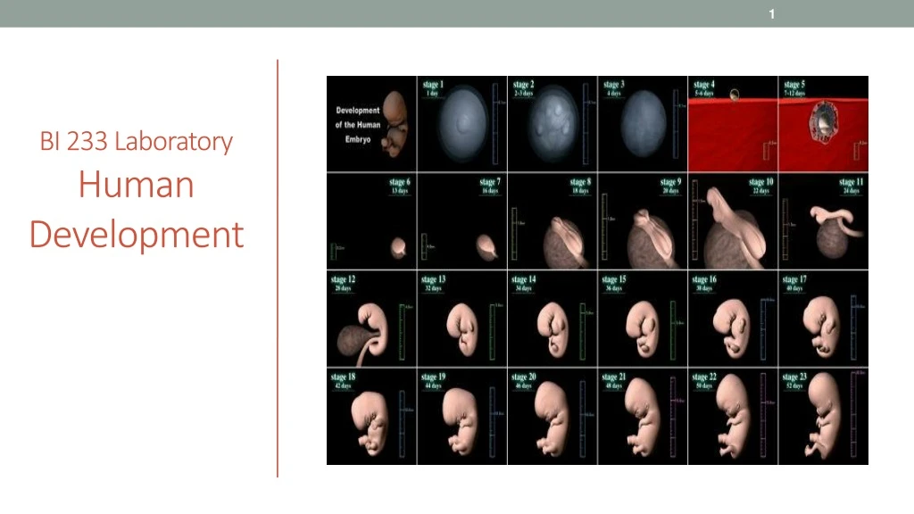
BI 233 Laboratory Human Development
360 likes | 375 Vues
Explore the incredible journey of human development, from spermatogenesis and oogenesis to blastocyst formation, implantation, and organogenesis. Learn about the fusion of gametes, cleavage divisions, and the formation of primary germ layers. Witness the miraculous process of creating a new life.

BI 233 Laboratory Human Development
E N D
Presentation Transcript
Spermatogenesis Involves:Mitosis, Meiosis and Spermiogenesis • Video on spermatogenesis
The Process of Oogenesis • AKA: ovum production – before birth • 2 million at birth • 11,000 die every month. • Accelerates at & after puberty. • Ends at menopause.
From Start of a Single Cell… Becomes the Being Containing Trillions • Fertilization • AKA: conception • Marks beginning of development • Differentiation • Formation of different types of cells • Selective changes in genetic activity • Genes are turned off, while others are turned on
Fertilization Defined • Fusion of two haploid gametes each containing 23 chromosomes • Produceszygotecontaining 46 chromosomes • 2000:1 size ration • Typically occurring in uterine tube. • Junction of ampulla and isthmus. • Sperm reach uterine tube from 30 – 120 min.
Activation of the Oocyte • ~ 200 sperm reach isthmus. (from 100 million) • Hyaluronidase released from acrosomal cap. • 1 sperm pushes through corona radiata. • Binding sperm triggers rupture of acrosome. • Acrosin digests path through zona pellucida. • Sperm and oocyte membranes fuse. • Activation of oocyte triggered. • Completion of meiosis II Fertilizing spermatozoon Second polar body
Pronucleus Formation Begins • Cortical reaction… • Inactivation of sperm receptors. • Completion of meiosis II, 2nd polar body forms. • Activation of metabolic enzymes. • mRNA activation for protein synthesis. • Sperm is absorbed into cytoplasm. • Female pronucleus develops. Female pronucleus Nucleus of fertilizing spermatozoon
Spindle Formation and Cleavage Preparation • Male pronucleusmigrates to center of cell. • Spindlefibers form. • Mark preparation for first cleavage division. Female pronucleus Male pronucleus
Fusion of Pronuclei = Amphimixis = Conception • Fusion of female and male pronuclei. • Moment of conception. • Now a zygotewith 46 chromosomes. • Fertilization is officially complete. • First cleavage division begins.
First Cleavage Forms Blastomeres • First cleavage division • Completion ~30 hours after fertilization. • Yields two daughter cells. • Each half the size of original zygote. • Known as blastomeres.
Cleavage and Blastocyst Formation The inner cell mass will form the embryo
Fertilization to Day 2 • Blastomeres • Identical cells produced by cleavagedivisions. • First division completed in ~30 hours. • Subsequent divisions occurring at intervals of 10-12 hours.
Day 3 to Blastocyst Formation • Morula (pre-embryo) • Solidball of cells, “mulberry” • Blastocyst • Hollow ball containing 3 parts. 1. Blastocoele 2. Trophoblast • Outer layer of cells. • Provides nutrients to embryo. • Takes part in placental formation. 3. Inner cell mass • Ultimately forms embryo.
Stages in Implantation: Days 6 and 7 • Implantation occurs seven days after fertilization. • Blastocyst attaches to endometrium. • Trophoblast will adhere to the endometrium. • Inner cell mass side facing the uterine wall. Uterine glands secrete glycogen-rich fluid
Stages in Implantation: Days 8and 9 • Trophoblast cells proliferate forms 2 layers. • Syncytiotrophoblast • Perimeter touching endometrium. • Cytotrophoblast • Interior of trophoblast retaining cell boundaries. • Creates syncytium • Syncytiotrophoblast erodes path. • Hyaluronidase enzyme • Ectopic pregnancy (aka tubal pregnancy) https://www.youtube.com/watch?v=bIdJOiXpp9g
Amniotic Cavity and Yolk Sac Formation • Implanted blastocyst covered by endometrial cells. • Chorionic villi • Extend into maternal blood filled lacunae. • Inner cell mass (ICM) • Organizes to become embryonic disc. • Ectoderm layer • Forms amniotic cavity. • Endoderm layer • Forms yolk sac. • Amnion • Cushions developing embryo and fetus. • Yolk sac primary nutrient source ICM.
Gastrulation:Migration of cell to interior forming layer between ectoderm and endoderm. • Creates primitive streak. • Raised dorsal groove establishes longitudinal axis of embryo. • Results in formation of 3rd germ layer. • Endoderm • Mesoderm • Ectoderm • Gastrulation sets the stage for organogenesis. • https://www.youtube.com/watch?v=3AOoikTEfeo
Primary Germ Layers • Serve as primitive tissues from which all body organs will be derived • Endoderm: • Epithelial lining of GI & lower respiratory tract • All ducts entering the GI tract • Lung, Liver, Pancreas • Urinary bladder • Ectoderm: • Brain & Nervous system • Epidermis • Lining of mouth, and anus • Sense organs such as eyes
Primary Germ Layers • Mesoderm: • Muscle • Bone • Cartilage • Cardiovascular • Dermis and hypodermis • Kidneys, • ovaries, testes • Lining of body cavities
Formation of Extraembryonic Membranes • Yolk sac • Blood cell formation. • Amnion • Contains amniotic fluid. • Allantois • Gives rise to urinary bladder. • Chorion • Transit system for blood and nutrients. • Chorionic villi • Becomes placenta.
Placentation • Body stalk • Yolk stalk • Decidua capsularis • Decidua basalis • Decidua parietalis • Umbilical cord
Placenta • Chorion of embryo and stratum functionalis layer of uterus. • Fully functional by 3rd month of pregnancy. • No union of maternal/fetal blood vessels. • Blood does not mix. • Diffusion of O2, nutrients, wastes, stores nutrients • produces hormones. • Barrier to microorganisms, except some viruses.
Fetal Circulation • Lungs are non-functional. • Oxygen and nutrients move from… • Maternal side of placenta. • To fetal bloodstream. • Via umbilical vein • CO₂ and wastes move from… • Fetal blood. • To placenta. • Via umbilical arteries
Fetal Circulation • Placental blood flows from umbilical cord • Through umbilical vein • To ductus venosus& inferior vena cava • To right atrium or through foramen ovale (shunt from R to L atrium) • From right ventricle flows to pulmonary trunk • Ductus arteriosus shunts blood flow from pulmonary trunk to aortic arch (bypassing lungs) • Flow continues via aorta to internal iliac arteries • Umbilical arteries branch from internal iliac arteries • Returning deoxygenated blood and waste to placenta and maternal circulation • Simulation of fetal circulation
The Endocrine Placenta - 6 • Human Chorionic Gonadotropin (hCG): • Maintenance of Corpus luteum (progesterone). • Prevents further uterine cycles. • Detected by some pregnancy tests • Human Placental Lactogen(hPL) • placental Prolactin • Prepares mammary glands for milk production. • Relaxin • Increases flexibility of pubis, cervix, vaginal canal. • Suppresses release of oxytocin until labor. • Progesterone • (later) Estrogen • Maintenance of endometrial lining.
Gestation…..Time Spent in Prenatal Development • First trimester • Embryological and early fetal development, rudiments of organs appear. • Second trimester • Development of organs and organ systems, fetus starts to look human. • Third trimester • Rapid fetal growth, deposition of adipose tissue, organs become functional.
Embryogenesis . • Begins shortly after gastrulation. ->PRIMARY GERM LAYERS • Embryo body begins to separate from embryonic disc. • Embryo body and internal organs start to form. • Folding, differential growth of disc form bulge projecting into amniotic cavity. • Head fold • Tail fold • First 12 weeks critical in establishing basis for organogenesis. • Process of organ formation. Week 2
First Trimester • 4 distinct processes… • Cleavage • Implantation • Placentation • Embryogenesis • Most dangerous period in prenatal life. • 40% of conceptions survive past first trimester.
The Mother: Second and Third Trimesters Growth of the Uterus and Fetus
Changes in Maternal System • ↑ Respiratory Rate, TV. • ↑ Maternal blood volume (up to 50%). • ↑ Maternal nutrient requirements (climb 10-30%). • ↑ Maternal GFR (increases by ~ 50%). • ↑ Size of uterus (7.5 cm, 30 g – 30 cm 1,100 g). • ↑ Mammary glands, breasts. • True labor. • False labor. • Labor contractions.
Development…..Gradual and Continuous Process From Fertilization to Maturity • Embryological Development • Occurs first 2 months after fertilization. • Study of events known as embryology. • Fetal Development • Begins at the ninth week. • Continues until birth.
Lifes Greatest Miracle • http://www.pbs.org/wgbh/nova/body/life-greatest-miracle.html • :41 • 6:50 • 15.10 • 26.48 • 40.24 • 43.49 • 50:17
Activities • Study and review all models of human development for structure identification. • Answer all associated questions in Survival Guide.
Works Cited Marieb, E. N. (2012). Essentials of human anatomy & physiology (6th ed.). Boston: Pearson Education, Inc. Marieb, E.N., Mitchell, S.J. & Smith, L.A. (2012). Human anatomy and physiology laboratory manual (10th ed.). Boston: Pearson Education, Inc. Martini, F., Nath, J. & Bartholomew, E.F. (2012). Fundamentals of anatomy & physiology (9th ed.). Boston: Pearson Education, Inc. McPhee, J. & Papadakis, M. (2012) Current medical diagnosis & treatment (51st ed.). New York: McGraw Hill. Patton, T. & Thibodeau, G. (2013). Anatomy & physiology (8th ed.). St. Louis:Mosby Elsevier. Saladin, K. S. (2012). Anatomy & physiology: The unity of form and function (6th ed.). New York: McGraw Hill. Tortora, G.J. & Derrickson, B.H. (2012). Principles of anatomy and physiology (13th ed.). Hoboken, NJ: Wiley