Download
1 / 41
1.16k likes | 8.86k Vues
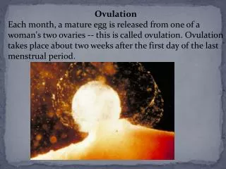
PHYSIOLOGY OF OVULATION. DR. ARCHANAVIKRAM ASST PROF DEPT OF OBG. DEFNITION. it is a process whereby a secondary oocyte is released from the ovary following rupture of a mature graffian follicle and becomes available for conception

E N D
PHYSIOLOGY OF OVULATION DR. ARCHANAVIKRAM ASST PROF DEPT OF OBG
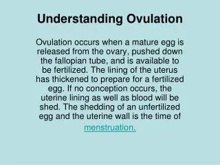
DEFNITION • it is a process whereby a secondary oocyte is released from the ovary following rupture of a mature graffian follicle and becomes available for conception Ovulation is the process of release of an oocyte from the ovarian follicle.
FOLLICULAR GROWTH AND ATRESIA • Germ cells • Primordial follicle • Cyclic maturation of the follicle (ovarian cycle) • Ovulation • Corpus luteum • Follicular atresia
At birth, the total number of primordial follicles is estimated to be about 2 million. The primary oocytes do not finish the first meiotic division until puberty-is reached. • At puberty, some 4,00,000 primary oocytes are left behind, the rest become atretic. During the entire reproductive period, some 400 are likely to ovulate.
44+ X+X oogenesis Oogonium 44+ X+X Primary oocyte 22+ X Secondary oocyte 22+ X First polar body 22+ X 22+ X Second polar body ovum
PRIMORDIAL FOLLICLE The primordial follicle consists of an oocyte, which is surrrounded by a-single layer-of flattened granulosa cells. The follicle measures about 0.03-0.05 mm. The oocyte (primitive ovum) measures about 18-24 diameter, nucleus 12 and nucleolus 6.
MORPHOLOGY OF THE OOCYTE It measures about 130 microns and the nucleus measures 20-25 microns. The radially arranged granulosa cells surrounding the oocyte- corona radiata . The oocyte is surrounded by an outer envelope called zona pellucida, a glycoprotein layer, secreted by the growing oocyte. The cytoplasm- vitellus contains nutritive yolk granules and is limited by a definite membrane called vitelline membrane.
The spherical nucleus is located near the centre of the cytoplasm. • The nucleolus is large with sparsely distributed chromatin. .
OVARIAN CYCLE • Definition • The development and maturation of a follicle, ovulation and formation of corpus luteum and its degeneration constitute an ovarian cycle. All these events occur within 4 weeks. • Thus, the ovarian cycle consists of: • Recruitment of groups of follicles. • Selection of dominant follicle and its maturation. • Ovulation. • Corpus luteum formation. • Demise of the corpus luteum.
Recruitment of groups of follicles(Preantral phase) The cohort of the growing follicles undergoes a process of development and differentiation which takes about 85 days. It is presumed that about 20 antral follicles (about 5-10 per ovary) proceed to develop in each cycle The initial recruitment - not under the control of any hormone. After a certain stage (2-5 mm in size)- FSH Unless the follicles are rescued by FSH at this stage, they undergo atresia.
Selection of a dominant follicle and its maturation The Graafian follicle is named after the Dutch physician and anatomist Reijnier de Graaf (1641- 1673). The development of antrum containing secondary or vesicular follicle from the solid primary follicle depends on FSH. There is accelerated growth of all the components of the follicles of the preantral phase. The fluid-filled space is formed amidst the granulosa cells. The spaces coalesce to form an antrurm.
Dominant follicle As early as day 5-7, one of the follicles out of so many becomes dominant and undergoes further maturation. The one with highest antral concentration of oestrogen and lowest androgen: oestrogen ratio and contain the maximum receptors for FSH, becomes the dominant follicle. The rest of the follicles become atretic by day 8
FSH induces LH receptors on the granulosa cells of the dominant follicle. LH receptor induction is essential for the midcycle LH surge to induce ovulation, luteinisation of the granulosa cells to from corpus luteum and secretion of progesterone.
Ovulation The dominant follicle, shortly before ovulation reaches the surface of the ovary. The cumulus becomes detached from the wall so that the ovum with the surrounding cells (corona radiata) floats freely in the liquor folliculi. The oocyte completes the first meiotic division with extrusion of the first polar body.The follicular wall near the ovarian surface becomes thinner. The stigma develops as a conical projection which penetrates the outer surface layer of the ovary.The stigma is soon closed by a plug of plasma
Causes singly or in combination. 1. Endocrinal LH surge Sustained peak level of oestrogen for 24-48 hours in the late follicular phase results in LH surge from the anterior pituitary (positive feedback effect). Effective LH surge persists for about 24 hours. • FSH rise • Preovulatory rise of 17--hydroxy progesterone facilitates the positive feedback action of oestrogen to induce FSH surge increase in plasminogen activator plasminogen plasmin helps lysis of the wall of the follicle.
2. Stretching factor It is more a passive stretching causing necrobiosis of the overlying tissue rather than rise in infrafollicular pressure which remains static at about 10-15 mmHg. 3. Contraction of the micromuscles in the theca externa and ovular stroma due to increased local prostaglandin secretion.
Corpus luteum After ovulation, the ruptured Graafian follicle develops into corpus luteum. Stage of proliferation: The collapsed walls of the empty follicle form convolutions. The granulosa cells undergo hypertrophy without multiplication. The cells are called granulosa lutein cells.
Stage of vascularisation Within 24 hours of rupture of the follicle, small capillaries grow into granulosa layer towards the lumen accompanied by lymphatics and fibroblasts. The sprouting vessels may rupture and bleed in the cavity. Stage of maturation By 4th day, the luteal cells have attained the maximum size.. Some theca lutein cells called ‘K’ cells invade the granulosa layer. The lutein cells develop lipid inclusion, giving the cells a distinctive yellowish colour.
Stage of Regression • On the day 22-23 of cycle, retrogression starts. The lutein cells atrophy and the corpus luteum becomes corpus albicans. Regression of corpus luteum is due to withdrawal of tonic LH support. • Hormones in relation to formation and maintenance of corpus luteum • FSH in presence of high level of oestrogen induces LH receptors in the granulosa cells of the dominant follicle. There after mid cycle LH surge causes luteinisation of the granulosa cells and progesterone secretion.
2. increased secretion of oestradiol and of 17--hydroxy progesterone is a prerequisite for adequate corpus luteum formation. 3. Low level of prolactin: The role of prolactin is not clear. Corpus luteum of pregnancy There is a surge of hyperplasia of all the layers between 23rd to 28th day due to chorionic gonadotrophin. hCG. The 'K' cells are also increased in number. It looks bright orange, later on becomes yellow and finally pale.
'messenger' to the pituitary gland providing a measure of the follicular growth LH surge follicular growth support a pregnancy
Hormone secretion Hormones-predominantly progesterone is secreted by the corpus luteum to support the endometrium of the luteal phase. There is also secretion of oestrogen, inhibin and relaxin.. This turn over of function from corpus luteum of pregnancy to placenta is called luteal-placental shift. This transition period continues from seven weeks to ten weeks.
FOLLICULAR ATRESIA • Atresia is a continued process which actually starts at 20th week of intrauterine life and ends at menopause. • Changes in the ovum occur first. • The ovum swells and undergoes hyaline and fatty degeneration. • Liquor folliculi is gradually absorbed with increased formation of hyaline tissue. • Eventually, the follicle collapses.
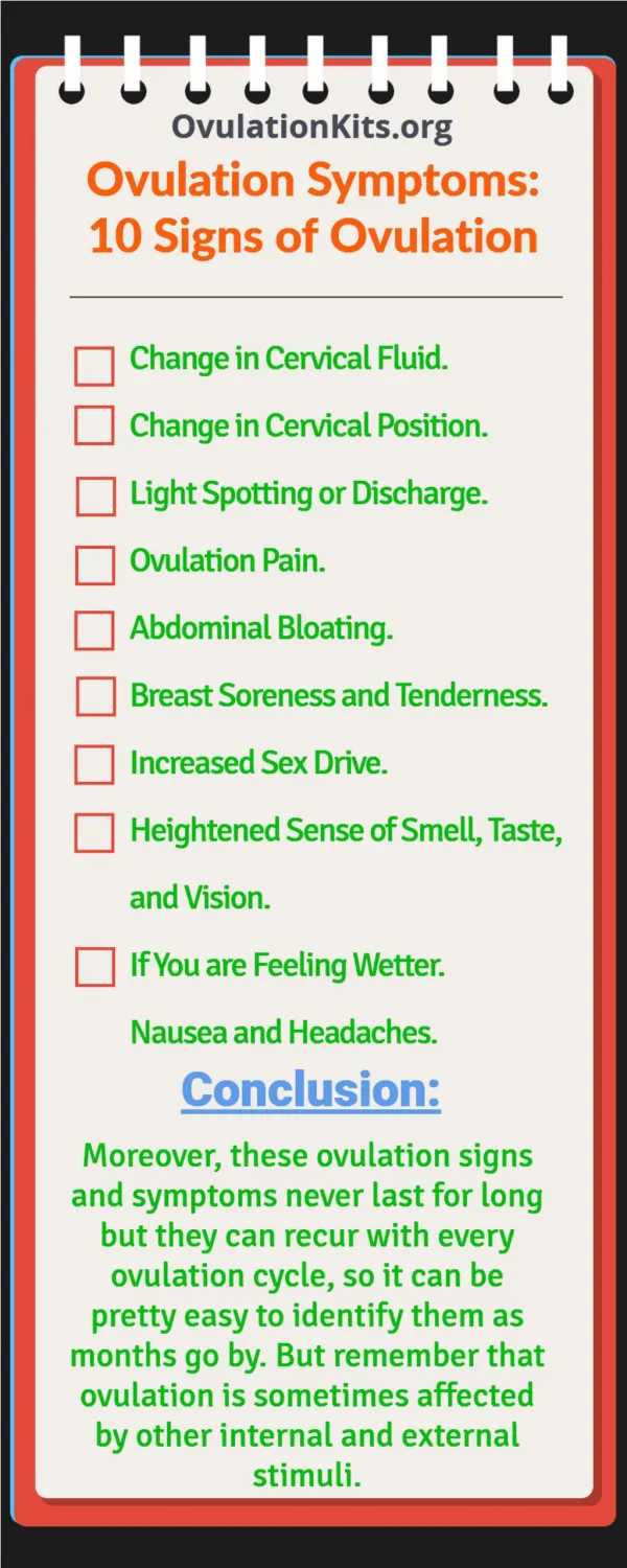
Indicators of ovulation • Determination of basal body temperature • Cervical mucus cycle • Endometrial biopsy • Estimation of hormone level in circulation- LH surge • Ultrasound examination of the follicle
ANOVULAR MENSTRUATION In an anovulatory cycle, the follicles grow without any selection of dominant follicle. The oestrogen is secreted in increasing amount. There may be imbalance between oestrogen and FSH or because of temporary unresponsiveness of the hypothalamus to the rising oestrogen, GnRH is suppressed no ovulation. The net effect is unopposed secretion of oestrogen till the follicles exist.
The endometrium remains in either proliferative or at times hyperplastic state. • There is inadequate structural stromal support and the endometrium remains fragile. • When the oestrogen level falls, there is asynchronus shedding of the endometrium and menstruation. • The bleeding may be heavy or prolonged and irregular.