Download
1 / 49
490 likes | 859 Vues
Relaxation of Pelvic Supports (Pelvic Organ Prolapse). Women’s Hospital, School of Medicine, Zhejiang University Jianhong Zhou 周坚红. INTRODUCTION. Pelvic organ prolapse(POP) is a common group of clinical conditions affecting millions of women Including anterior and posterior vaginal prolapse

E N D
Relaxation of Pelvic Supports(Pelvic Organ Prolapse) Women’s Hospital, School of Medicine, Zhejiang University Jianhong Zhou 周坚红
INTRODUCTION • Pelvic organ prolapse(POP) is a common group of clinical conditions affecting millions of women • Including anterior and posterior vaginal prolapse • Uterine prolapse and enterocele
CLINICAL IMPORTANCE • The prevalence of POP increases with age • The lifetime risk that a woman in the USA will have surgery for POP or urinary incontinence is 11%, with up to 1/3 of surgeries representing repeat procedures. • The direct cost of prolapse surgery is greater than $1 billion per year
CLINICAL IMPORTANCE • Surgically treated prolapse represents the severe end of the clinical spectrum • For the vast majority of asymptomatic women with physical findings of prolapse, no treatment is indicated
Anatomy of pelvic floor • To understand the pathophysiology of POP • Some knowledge of normal vaginal support is needed
The female pelvic floor serves to aid in the function of the lower urinary system, genital tract and rectum. The female pelvic floor is composed of voluntary muscle, fascia and condensations of fascia called ligaments that all work together to offer support and function to the organs that exit through the female pelvis
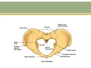
an empty pelvis looking from above. At the back is the sacrum (tailbone) and the front is the pubic bones
Exiting the female pelvis from the front to the back, is the urethra, vagina and anus. • The anus is surrounded by the anal sphincter which is connected to the sacrum behind and the perineum muscles in front. • The perineum stabilizes the lower pelvic floor. The perineum is stabilized to the pelvic side-wall by the transverse perineum muscles.
The urogenital diaphram offers further support to the lower urethra and vagina and is stabilized to the pubic bones and the perineum
The pelvic floor muscles form a sling around the lower pelvic outlet and when contracted offers support to the lower bowel, the genital tract and the lower urinary tract. These muscles arise from the white line on lateral pelvic side-wall and interdigitate with each other and the anococcygeal raphe in the centre.
The rectum lies on the pelvic floor and joins to the anus as it passes through the pelvic floor.
The rectum is stopped from bulging forward into the vagina by the rectovaginal fascia. • Women with rectoceles (rectum bulging forward into the vagina) have defects in this fascia.
The mid-vagina rest on the rectovaginal fascia and is separated from the bladder and urethra by the anterior vaginal wall fascia (pubocervical). • Women with cystoceles (bladder prolapse) have a defect in this fascia.
The bladder and urethra are separated from the vagina by the pubocervical fascia. Intact fascia stops the bladder from bulging down into the vagina.
The top of the fascia on the anterior and posterior vaginal wall connect with cervix at the top of the vagina. • The cervix is the very firm lower portion of the uterus and acts to stabilize the fascia of the female pelvis.
The cervix is supported to the lateral pelvic side-wall by the strong cardinal ligaments.
The cervix is also supported by the strong uterosacral ligaments that run from the sacrum (tailbone) to the cervix. • Women with upper vaginal prolapse (uterine prolapse, vault prolapse or enteroceles) have defects in the uterosacral and / or cardinal ligaments that allows the prolapse to occur
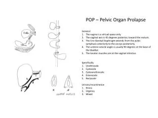
Women with upper vagina prolapse have a defect of the cardinal and uterosacral defects. • Cystoceles ——weakness of the pubocervical fascia. • Rectoceles —— tears or weakness of the rectovaginal fascia. • The cervix and perineum are the centre points for stabilization of the pelvic floor.
LEVEL 1 Cardinaluterosacral ligament complex To arcus tendineous fascia complex LEVEL 2 To arcus tendineous rectovaginalis LEVEL 3 Obturator foramen Perineal membrane Superficial transverse Perineal muscle
Delancey’s three levels of support are now accepted worldwide • Level 1: The cardinal-uterosacral ligament complex provides apical attachment of the uterus and vaginal vault to the bony sacrum. Uterine prolapse occurs when the cardinal-uterosacral ligament complex breaks or is attenuated. • Level 2: The arcus tendineous fascia pelvis and the fascia overlying the levator ani muscles provide support to the middle part of the vagina. • Level 3: The urogenital diaphragm and the perineal body provide support to the lower part of the vagina.
Potential Risk Factors for POP • Predispose • Incite • Promote • Decompensate
Predispose • Genetic (congenital or hereditary) • Race: White African-American • Gender: Female> Male
Incite • Pregnancy and delivery-birth trauma • Surgery such as hysterectomyfor prolapse(11.6%/1.8%) • Myopathy • Neuropathy
Promote • Obesity(40%--75%) • Smoking • Pulmonary disease (chronic coughing) • Constipation (chronic straining) • Recreational or occupational activities(frequent or heavy lifting)
Decompensate • Aging • Menopause • Neuropathy • Myopathy • Debilitation • Medication
Symptoms • Not specific to different compartments of prolapse but may reflect the overall stage of prolapse at its most advanced site • Pelvic pressure or heaviness • Vaginal bulge • Pelvic pain was not associated with prolapse, and women with more advanced prolapse had less back pain than women with mild prolapse
Symptoms • Women with urethral incompetence are continent only because the prolapse causes urethral kinking and obstruction • Potential, masked, latent, or occult stress incontinence because women do not have symptoms of incontinence as long as the prolapse is untreated.
Physical Examination • Focuses on the pelvic examination • Beginning with a careful inspection of the vulva and vagina to identify erosions, ulcerations, or other lesions • Suspicious lesions should be biopsied • The extent of prolapse should be systematically assessed
Pelvic Organ Prolapse Quantification system • Measures 9 locations on the vagina and vulva in centimeters relative to the hymen • These 9 locations are used to assign a stage (from 0 to IV) of prolapse at its most advanced site • More detailed than necessary for clinical care
2 most important advantagesover previous grading systems 1) The standardized technique with quantitative measurements at straining relative to a constant landmark, the hymen 2) Prolapse assessment at multiple vaginal sites
The five stages of prolapse • Stage 0: No prolapse • Stage I: The most distal portion of the prolapse is >1 cm above the level of the hymen • Stage II: The most distal portion of the prolapse is ≤1cm proximal or distal to the hymen • Stage III: The most distal portion of the prolapse is >1 cm below the hymen but protrudes no further than 2 cm less than the total length of the vagina • Stage IV: Complete eversion of the vagina
Baden-Walker Halfway system • Most clinicians utilize this system • It records the extend of prolapse using a four-point using the hymen as a fixed point of reference 1: descensus halfway to the hymen 2: descensus to the hymen 3: descensus halfway past the hymen 4: maximum descent
THERAPEUTIC APPROACH • Observation • Pelvic floor rehabilitation • Pessary use • Surgery
Indications for Treatment • Depends on symptom severity and severity of prolapse • Patient’s general health and activity • The correlation between many pelvic symptoms and the extent of prolapse is weak
Observation • Appropriate for women whose symptoms are not sufficiently bothersome to warrant active intervention • There is virtually no indication for treatment, particularly surgery, for women with asymptomatic prolapse • The old adage “You can’t make an asymptomatic patient better; you can only make her worse” was never more true than it is for prolapse.
Nonsurgical Management • Adjunct therapy to address concomitant symptoms, pelvic floor muscle training, and pessaries. • Nonsurgical management • decrease the frequency and severity of symptoms • delay or avoid surgery • potentially prevent worsening the prolapse
Pelvic Floor Muscle Training • Be designed to increase the strength and endurance of the pelvic muscles • Thereby improving support to the pelvic organs
Pessaries • To decrease symptom frequency and severity • Delay or avoid surgery • Potentially prevent worsening of prolapse • Relative contraindication to pessary use is persistent vagina erosions
Surgical Management • Aim of surgery is to relieve or improve prolapse symptoms • Symptoms associated with the lower urinary and gastrointestinal tracts. • In some women, this means an attempt to restore normal vaginal anatomy and maintain or improve sexual function. • In others, an obliterative approach is more appropriate and still yields the desired result of symptom relief.
Surgical procedures for POP • Hysterectomy for uterine prolapse • Anterior repair, paravaginal repair for cystocele • Posterior repair for rectocele • Enterocele repair • Vaginal vault suspension • Perineorrhaphy for relaxed vaginal outlet
Treat patients individually • The best choice is the first choice • The first choice is the best choice
THANK YOU FOR YOUR ATTENTION Additional educational resources-Resources for health professionals Conservative management of pelvic organ prolapse in women. www.cochrane.org/reviews/en/ab003882.html Mechanical devices for pelvic organ prolapse in women. www.cochrane.org/reviews/en/ab004010.html Surgical management of pelvic organ prolapse in women. www.cochrane.org/reviews/en/ab004014.html BMJ Clinical Evidence. Genital prolapse in women. www.clinicalevidence.com/ceweb/conditions/woh/0817/0817.jsp


![get [PDF] Download Pelvic Organ Prolapse: The Silent Epidemic download](https://cdn7.slideserve.com/12583059/pelvic-organ-prolapse-the-silent-epidemic-dt.jpg)
