Download
1 / 46
460 likes | 574 Vues
Learn about vesicle transport processes - exocytosis and endocytosis, key mechanisms in transporting macromolecules across the cell membrane.

E N D
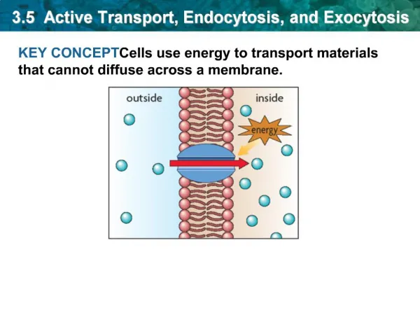
Vesicle Transport • In diffusion, dissolved particles (solutes) move down a concentration gradient. • Some molecules or particles are just too large to pass through the plasma membrane or to move through a transport protein. • Cells use two other active transport processes to move these macromolecules (large molecules) into or out of the cell. • Vesicles are the bodies in the cytoplasm which help to move macromolecules or large particles across the plasma membrane.
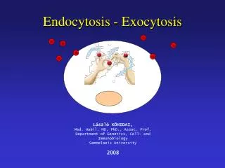
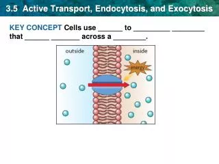
There are two types of vesicle transport, endocytosis and exocytosis. • Both processes are active transport processes, requiring energy.
Endocytosis: {Endo (within) cytosis (cell)} : The process by which a cell moves large amounts of material, or non-dissolved particles, into its cytoplasm from the outsideenvironment. Exocytosis: {Exo (exit) cytosis (cell)} : The process by which large amounts of material, or large non-dissolved particles, are moved from the cell’s cytoplasm to the outsideenvironment.
Exocytosis • Exocytosis describes the process of vesicles fusing with the plasma membrane and releasing their contents to the outside of the cell. • Exocytosis occurs when a cell produces substances for export, such as a protein, or when the cell is getting rid of a waste product or a toxin. • Newly made membrane proteins and membrane lipids are moved on top the plasma membrane by exocytosis.
In exocytosis, membrane – bound secretory vesicles are carried to the cell membrane, and their contents are secreted into the extracellular environment. • The secretion is possible because the vesicle transiently fuses with the outer cell membrane. • Exocytosis is also a mechanism by which cells are able to insert membrane proteins (such as ion channels and cell surface receptors), lipids, and other components into the cell membrane.
Exocytotic Vesicles • Vesicles containing protein products are typically derived from an organelle called the Golgi apparatus or Golgi complex. • Proteins and lipids synthesized in the endoplasmic reticulum are sent to Golgi complexes for modification and sorting. • Once processed, the products are contained within secretory vesicles, which bud from the Golgi apparatus. • Some vesicles are formed from early endosomes, which are membrane sacs found in the cytoplasm. • These endosomes sort the internalized material (proteins, lipids, microbes, etc.) and direct the substances to their proper destinations.
Types of Exocytosis • There are three common pathways of exocytosis. • Constitutive exocytosis • Regulated exocytosis • Lysosome pathway
Constitutive exocytosis • Constitutive exocytosis involves the regular secretion of molecules that is performed by all cells. • This pathway serves to deliver membrane proteins and lipids to the cell's surface and to expel substances to the cell's exterior.
Regulated exocytosis • Regulated exocytosis relies on the presence of extracellular signals for the expulsion of materials within vesicles. • Regulated exocytosis occurs commonly in secretory cells and not in all cell types. • Secretory cells store products such as hormones, neurotransmitters, and digestive enzymes that are released only when triggered by extracellular signals. • Secretory vesicles are not incorporated into the cell membrane, but fuse only long enough to release their contents. • Once the delivery has been made, the vesicles reform and return to the cytoplasm.
Lysosome pathway • A third pathway for exocytosis in cells involves lysosomes. • These organelles contain acid hydrolase enzymes that break down waste materials, microbes, and cellular debris. • Lysosomes carry their digested material to the cell membrane where they fuse with the membrane and release their contents into the extracellular matrix.
Steps of Exocytosis • Exocytosis occurs in four steps in constitutive exocytosis and in five steps in regulated exocytosis. These steps include vesicle trafficking, tethering, docking, priming, and fusing. • Trafficking- Vesicles are transported to the cell membrane along microtubules of the cytoskeleton. Movement of the vesicles is powered by the motor proteins kinesins, dyneins, and myosins. • Tethering- Upon reaching the cell membrane, the vesicle becomes linked to and pulled into contact with the cell membrane. • Docking- Docking involves the attachment of the vesicle membrane with the cell membrane. The phospholipidbilayers of the vesicle membrane and cell membrane begin to merge.
Priming- Priming occurs in regulated exocytosis and not in constitutive exocytosis. This step involves specific modifications that must happen in certain cell membrane molecules for exocytosis to occur. These modifications are required for signaling processes that trigger exocytosis to take place. • Fusion- There are two types of fusion that can take place in exocytosis. • In complete fusion, the vesicle membrane fully fuses with the cell membrane. The energy required to separate and fuse the lipid membranes comes from ATP. The fusion of the membranes creates a fusion pore, which allows the contents of the vesicle to be expelled as the vesicle becomes part of the cell membrane. • In kiss-and-run fusion, the vesicle temporarily fuses with the cell membrane long enough to create a fusion pore and release its contents to the exterior of the cell. The vesicle then pulls away from the cell membrane and reforms before returning to the interior of the cell.
Exocytosis Examples • Exocytosis is used by a number of cells in the body as a means of transporting proteins and cell to cell communication. In the pancreas, small clusters of cells called islets of Langerhans produce the hormones insulin and glucagon. • These hormones are stored in secretory granules and released by exocytosis when signals are received. • When glucose concentration in the blood is too high, insulin is released from islet beta cells causing cells and tissues to take up glucose from the blood. • When glucose concentrations are low, glucagon is secreted from islet alpha cells. • This causes the liver to convert stored glycogen to glucose. • Glucose is then released into the blood causing blood-glucose levels to rise. • In addition to hormones, the pancreas also secretes digestive enzymes (proteases, lipases, amylases) by exocytosis.
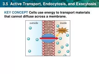
Endocytosis • Endocytosis is the case when a molecule causes the cell membrane to bulge inward, forming avesicle. • Endocytosis is a form of active transport in which a cell transports molecules (such as proteins) into the cell by engulfing them in an energy-using process. • The three types of endocytosisare: • Receptor mediated endocytosis • Phagocytosis • Pinocytosis
Receptor mediated endocytosis • Receptor-mediated endocytosis (RME), also called clathrin-mediated endocytosis, is a process by which cells absorb metabolites, hormones, other proteins – and in some cases viruses – by the inward budding of plasma membrane vesicles containing proteins with receptor sites specific to the molecules being absorbed. • Endocytoticmechanism in which specific molecules are ingested into the cell. • The specificity results from a receptor-ligand interaction.
Receptor mediated endocytosis • The protein Clatherin coats the inside of the membrane in the area of the pit. • Receptors on the plasma membrane of the target tissue will specifically bind to ligands on the outside of the cell. • An endocytotic process occurs and the ligand isingested. • Two chemical compounds called Pitstop 1 and Pitstop 2, selective clathrin inhibitors, can interfere with the pathogenic activity, and thus protect the cells against invasion.
Clathrin • Clathrin is a protein that plays a major role in the formation of coated vesicles. • Clathrin was first isolated and named by Barbara Pearse in 1976. • It forms a triskelion shape composed of three clathrin heavy chains and three light chains. • They form a polyhedral lattice that surrounds the vesicle.
Steps Of ReceptorMediated Endocytosis Step1: 1 Extracellularfluid Ligand binds to membranereceptor. Receptor Intracellularfluid
Step2 Extracellularfluid Ligand binds to membrane receptor. 1 Receptor-ligand migrates to clathrin-coatedpit. 2 Clathrin- coatedpit Receptor Clathrin Intracellularfluid
Step3 Extracellularfluid Ligand binds to membrane receptor. 1 Receptor-ligand migrates to clathrin-coatedpit. 2 Clathrin- coatedpit Endocytosis 3 Receptor Clathrin Intracellularfluid
Step4 Extracellularfluid Ligand binds to membranereceptor. 1 Receptor-ligand migrates to clathrin-coated pit. 2 Clathrin- coatedpit Endocytosis 3 Receptor Clathrin Vesicleloses clathrincoat. 4 Intracellularfluid
Step5 Extracellularfluid Ligand binds to membrane receptor. 1 Receptor-ligand migrates to clathrin-coated pit. 2 Clathrin- coatedpit 3 Endocytosis Receptor Clathrin Vesicleloses clathrincoat. 4 Receptors andligands separate. 5 Intracellularfluid Endosome
Step6 Extracellularfluid Ligand binds to membranereceptor. 1 Receptor-ligand migrates to clathrin-coated pit. 2 Clathrin- coatedpit Endocytosis 3 Receptor Clathrin Vesicleloses clathrincoat. 4 Receptors andligands separate. 5 To lysosomeor Golgicomplex 6 Intracellularfluid Ligands go to lysosomes or Golgi forprocessing. Endosome
Step7 Extracellularfluid Ligand binds to membrane receptor. 1 Receptor-ligand migrates to clathrin-coated pit. 2 Clathrin- coatedpit Endocytosis 3 Receptor Clathrin Transport vesicle with receptorsmoves to the cellmembrane. 7 Vesicleloses clathrincoat. 4 Receptors andligands separate. 5 To lysosomeor Golgicomplex Intracellularfluid Ligands go to lysosomes or Golgi forprocessing. 6 Endosome
Step8 Extracellularfluid Ligand binds to membrane receptor. 1 Receptor-ligand migrates to clathrin-coated pit. 2 Transport vesicle and cell membrane fuse (membrane recycling). 8 Clathrin- coatedpit 3 Endocytosis Receptor Clathrin Transport vesicle with receptors moves to the cellmembrane. 7 Vesicleloses clathrincoat. 4 Receptors andligands separate. 5 To lysosomeor Golgicomplex Intracellularfluid Ligands go tolysosomes or Golgi forprocessing. 6 Endosome
LastStep Extracellularfluid Ligand binds to membranereceptor. 1 Exocytosis 9 Receptor-ligand migrates to clathrin-coated pit. 2 Transport vesicle and cellmembrane fuse (membrane recycling). 8 Clathrin- coatedpit Endocytosis 3 Receptor Clathrin Transport vesicle with receptors moves to the cellmembrane. 7 Vesicleloses clathrincoat. 4 Receptors andligands separate. 5 To lysosomeor Golgicomplex Intracellularfluid Ligands go tolysosomes or Golgi forprocessing. 6 Endosome
Diagrammaticallyexplanation 1 Ligand binds tomembranereceptor. Extracellularfluid 9 Exocytosis Receptor-ligand migrates to clathrin-coated pit. Clathrin- 2 Transport vesicle and cellmembrane fuse (membrane recycling). 8 Endocytosis 3 coatedpit Receptor Clathrin Transport vesicle with receptors moves to the cellmembrane. 7 Vesicleloses clathrincoat. 4 Receptors andligands separate. 5 To lysosomeor Golgicomplex Intracellularfluid Ligands go to lysosomes or Golgi forprocessing. 6 Endosome
Uptake ofcholesterol • The uptake of cholesterolby mammalian cells has provided a key model receptor-mediated endocytosisat for understanding the molecular level. • Cholesterol is transported through the bloodstream in the form of lipoprotein particles, the most common of which is called low-density lipoprotein, orLDL. • The uptake of LDL by mammalian cells requires the binding of LDL to a specific cell surface receptor that is concentrated in clathrin-coated pits and internalized by endocytosis.
What isphagocytosis It is defined as ingestion of particlesof >0.5µm bycells.
Phagocytosisis one type of endocytosisthat occurs when a cell uses its membrane to bring non-dissolved particles orsolid particles into its cytoplasm. • In phagocytosis, the cell extends finger-like projections of its cell membrane, called pseudopods, around a piece of solid material outside of thecell. • The pseudopods that surround the solid object eventually join to form a vacuole with in the cells cytoplasm. • The cell then releases chemicals into the vacuole. • The chemicals digest the solid particle into smaller particles that maybe used for energy or buildingmaterial. • Phagocytosis is also often called “celleating”.
Who discoveredphagocytosis ? • Ellie Ilya Metchnikoff discovered it in 1882. • Received Nobel prize for the samein 1906 • Carl FredrichClaus coined theterm Phagocytosis.
Examples WBC remove bacteria and celldebris.
Cells involved - Professional phagocytes- have specific receptors1.Neutrophils 2.Monocytes 3.Macrophages 4.Dendritic cells5.Mast cells 6.Eosinophils
NoNProfessioNals Phagocytosisis not their main function , do not have specific receptors. • Lymphocytes • NK and LGL cells (Large Granularlymphocytes) • Epithelial cells Endothelialcells Fibroblasts
How does itwork Entry of thepathogen Phagosomes Phagolysosomes Recruitment of phagocytes &inflammation
Pinocytosis • Pinocytosis is another form of endocytosis in which small particles are brought into the cell, forming an invagination and then suspended with in small vesicles and then it breaks down the particles. • Pinocytosis works just like phagocytosis but the only difference other hand, pinocytosis is when the cell engulfs already-dissolved or broken-downfood. • Pinocytosis is also known as “cell drinking”.
Example Human egg cell taking up nutrients from surroundingcells.