Download
1 / 63
820 likes | 1.64k Vues
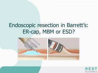
Endoscopic resection in Barrett’s: ER-cap, MBM or ESD?. In general: Unifocal, limited size (< 2cm); Mucosal lesions; Well / moderately differentiated neoplasia: How do I know it’s mucosal? Endoscopic appearance; Endoscopic ultrasound ER as a diagnostic procedure.

E N D
In general: Unifocal, limited size (< 2cm); Mucosal lesions; Well / moderately differentiated neoplasia: How do I know it’s mucosal? Endoscopic appearance; Endoscopic ultrasound ER as a diagnostic procedure Indications for ER in Barrett’s
Histologicalevaluation ER specimen: The most important step of the diagnosticwork-up.
Treatment concept and considerations ER RFA Surgery superficial sm invasion deep sm invasion IMC LGIN HGIN
Treatment concept and considerations All visible lesions or suspicious areas require endoscopic resection!
Submucosal liftingLifting sign (Adrenalin 1:20.000) Kato type 1: complete/soft Kato type 2: complete/hard Kato type 3: incomplete Kato type 4: “non-lifting sign” (Kato et al, Endoscopy 2001, 33: 568-573)
Start with injecting at the distal margin; In tubular esophagus: lesion at 6 o’clock position; In distal esophagus: retroflex; Insert the needle tangential to esophageal wall; Start fluid injection just before insertion; Start injecting at the edges; Avoid injection through lesion. Submucosal liftingInjection Technique
X X Submucosal liftingType 3: Incomplete lifting All sm1-sm3
X Submucosal liftingType 4: “no lifting” sign All sm3
ER-cap vs.MBM techniqueRandomised trial Pouw et al. Gastrointest. Endosc. 2011
ER-cap vs.MBM techniqueRandomised trial • Piecemeal ER with MBM is faster and cheaper than with the ER-cap technique; • MBM may be associated with fewer complications; • MBM results in significantly smaller sized resections; • MBM may, therefore, be more suited for resection of flat lesions with a low risk of submucosal invasion; • The ER-cap technique may be preferred for ER of elevated and nodular lesions. Pouw et al. Gastrointest. Endosc. 2011
Hard caps, straight or oblique, Ø: 12.8-14.8 mm. Hard, wide caps, straight or oblique, Ø: 16.1 mm. Large flexible oblique caps, Ø: 18 mm. ER-caps
Oblique caps for most lesions. Straight caps only for lesions that can be approached en-face (e.g. greater curvature stomach). Size of the cap = size of specimen. Size of the cap = depth of resection. ER-capsGeneral rules General rule Largecaliber cap foren-blocresections. Standard cap forpiecemealresections. Matzusaki et al Gastrointest. Endosc.2003
Different diameters to fit differently sized endoscopes. Choose right cap for endoscope. Fix with water resistant tape. Introduction hard cap can be difficult. ER-caps
Crescent shaped, single use ER snare ER-cap procedureSnares
Snare is placed in the distal ridge inside the cap. Position cap at an area with normal mucosa. Seal (not fill) the cap with gentle suction. Open snare slowly, keep tip at 6-9” position. Ideally the snare should open clockwise. ER-cap procedureSnare placement
Location lesion prior to resectionPosition the lesion at 6 o’clock
Location lesion prior to resectionPosition the lesion at 6 o’clock
Rotate the endoscope to position the lesion at the 6 o’clock position
Test suction prior to placement of snare may be useful. Amount of aspiration of mucosa in cap determines size resected specimen. En-bloc resections: go for complete “red-out”, be more conservative for piecemeal procedures. Tighten snare quickly until resistance is encountered. Suck and catch
Resect outside the cap. Hold the snare by yourself! Re-open 1-2 mm, inflate esophagus and shake specimen. Pre-coagulation for 1-2 seconds. Use either coagulation or Endocut for further resection. Erbe ICC 200: pre-coagulation: 1-2 sec 45 Watt. transsection: Endocut 120 Watt, effect 3. Resection
Multi-Band Mucosectomy (DuetteR) • Modified variceal band ligator: • Widened threading channel of the cranking device from 2 to 3.2 mm, allowing introduction of 7F accessories alongside the thread. • 7F accessories: • Not only a snare, but also an APC probe, spraying catheter, clipping device or hot biopsy forceps.
Multi-Band Mucosectomy (DuetteR) • Transparent cap with 6 rubber bands, to create a pseudopolyp; • Hexagonal snare for resection using electrocautery, reusable due to shape stability.
A pseudopolyp is created by suctioning mucosa into the cap, and releasing a rubber band; The snare should be placed below the rubber band; The snare should be closed tighter than with the cap-technique; Use pure coagulation (ICC 200, 45 W), usually the polyp is resected after 1 to 2 seconds. Multi-Band Mucosectomy (DuetteR)
Do not use the irrigation adapter of the set to puncture and widen the opening of the cranking device! Fix the Duette barrel properly. During fixing of the barrel, take care of the outer covering of the scope which may be damaged Align trigger cords preferrably at 6-12 o´clock position (Olympus scope). Mounting of the EMR device
Technical difficultiesdecreased visibility due to bands and wires Release some bands if you expect to perform en-bloc resection.
Therapeutic endoscope preferred due to better suction ability; Clean the area for ER of any secretion; Marking of the lesion by APC: 2-5 mm outside the lesion’s margins Tip of snare: Erbe ICC 200, 45 Watt APC: Erbe ICC 200 + APC 300, 40 Watt. Suck out all fluids from the stomach !!! Before ER
Try to remove the most involved area in a single piece, usually in the first resection; The lateral edges of each resection are the most superficial so avoid cutting through the area where you expect the deepest infiltration. Piecemeal ERWhat to target first?
Piecemeal ERSubsequent resections • Target the second resection area; • Variables: • Relative position of the cap to the resection wound; • Size of the cap; • Amount of aspiration of mucosa into the cap. • ER-cap: repeat lifting before every subsequent resection; • Always perform a test suction (and prior to placement of snare when using ER-cap).
Safety and efficacy of multiband mucosectomy in 1060 resections in Barrett's esophagus Herrero A. et al. Endoscopy 2011;43:177 Soehendra N. et al. GIE 2008;
Enables widespread ER; Technically more challenging than en-bloc resections; Bigger risk of complications; Recurrences appear more often after piecemeal procedures. En-bloc vs. Piecemeal ERpiecemeal resection
To prevent perforation do not suck too much at the EG junction and in a hiatal hernia. Avoid "red out phenomenon“ (suction of the tissue all the way to the lens). Practical aspectsTips & Tricks I
Torque the scope to appropriately locate the lesion according to the distal opening of the working channel to facilitate snaring. Avoid touching the pseudopolyp: do not move the barrel beyond the pseudopolyp extend and open the snare beyond the pseudopolyp pull the opened snare back and place it around the pseudopolyp Practical aspectsTips & Tricks II
Avoid too much overlap to prevent perforation; Less than 25% overlap is acceptable: resection is usually less deep at the overlapping lateral margins than at the centre of the resection; Avoid residual Barrett’s tissue between adjacent resections; Be liberal with submucosal injection in the resection area. Overlap vs. irradical resection
Useful in case of (very) small residual isles of target tissue between subsequent resections or between markings and the outer wound margin. Additional ablation residual tissueAPC 60-80 Watt
Always retrieve specimens for histopathological evaluation. This can be done at the end of the procedure. Retrieval: aspirate in cap (single specimen) or foreign body basket (multiple specimens). Pin specimen on paraffin/cork before fixation. Retrieval specimens
ESD in early Barrett's cancer ? Deprez P. et al. DDW 2010