Download
1 / 18
250 likes | 884 Vues
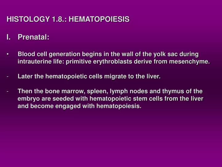
HISTOLOGY 1.8.: HEMATOPOIESIS Prenatal: Blood cell generation begins in the wall of the yolk sac during intrauterine life: primitive erythroblasts derive from mesenchyme. Later the hematopoietic cells migrate to the liver. Then the bone marrow, spleen, lymph nodes and thymus of the

E N D
HISTOLOGY 1.8.: HEMATOPOIESIS • Prenatal: • Blood cell generation begins in the wall of the yolk sac during • intrauterine life: primitive erythroblasts derive from mesenchyme. • Later the hematopoietic cells migrate to the liver. • Then the bone marrow, spleen, lymph nodes and thymus of the • embryo are seeded with hematopoietic stem cells from the liver • and become engaged with hematopoiesis.
II. Postnatal hematopoiesis The major site of hematopoiesis is the bone marrow, it also persists in the liver and spleen for a few weeks after birth (potential is retained). Early in life all the marrow is active, with age the demand for blood cells decreases: red marrow is replaced by resting yellow marrow. Red bone marrow is present in the adult animal: sternum vertebrae ribs skull pelvis epiphyses of long bones
Structure of the bone marrow The hematopoietic compartment consists of irregular anastomosing cords that lie between vascular sinuses. The marrow lacks lymphatic vessels Innervation: vasomotor nerves around the blood vessels Bone marrow smear Bone marrow in sternum Hematopoietic compartment
Blood cells are produced in the hematopoietic compartment and reach the bloodstream by crossing the wall of vascular sinuses. Adventitial reticular cells: meshwork that support hematopoietic cells and provide special microenvironment that influence the development of the various stem cells. They may fill with fat and transform into adipose cells when hematopoiesis decreases. Marrow hematopoiesis: is most active in areas close to the bone. Erythropoietic cells and megakaryocytes are close to vascular sinuses. Granulopoietic cells are deep within the cords, away from the vascular sinuses.
Hematopoietic stem cells: Undifferentiated cells are generally larger than mature cells, they have large euchromatic nuclei (large nuclear-to-cytoplasmic ratio). The marrow contains several types of self-replicating stem cells. Their morphology resembles that of lymphocytes, but their proliferating capacity differs: pluripotent multipotent stem cells unipotent Embryonic yolk sac, fetal liver,spleen, bone marrow: primitive stem cells (pluripotent) Lymphoid stem cells myeloid stem cells (multipotent) Lymphocytes erythrocytic granulocytic monocytic megakaryocytic (They are all unipotent cell types)
CFU-E (Colony-forming unit-erythrocyte) (Rubriblast) 16-22 mm (Metarubricyte) 12-15 mm (Rubricyte) 14-18 mm (Prorubricyte) 10-12 mm
Erythron: mass of circulating erythrocytes + marrow erythropoietic tissue Efficacy of erythropoiesis in a dog: 1 million/second Erythroblastic islands: group of erythrocytic cells within the bone marrow organized around a macrophage (more mature ones at the periphery).
1. Rubriblast (arrow) largest, with deep blue cytoplasm and round euchromatic nucleus 2. Prorubricyte (arrowhead) Similar, but smaller, no nucleoli 4. Metarubricyte: the smallest nucleated erythrocyte with picnotic nucleus 4. 3. 3. Basophilic rubricyte: nucleus with clumped chromatin
Reticulocyte Mature mammalian erythrocytes Erythrocyte kinetics: Development of rubriblasts to mature erythrocytes: 5-7 days Reticulocytes: normally 1-2 days maturation within the bone marrow Regulation of erythrocyte formation: cellular and humoral factors erythropoietin: key-molecule produced in the kidney
Granulopoiesis Occurs in clusters away from the vascular sinuses in the midportion of the hematopoietic compartment of the marrow CFU-GM: colony-forming units-granulocyte-monocyte (bipotent) Neutrophil and /or monocyte progenitor cells 15-18 mm 10-15 mm 18-22 mm
Myeloblast: ovoid or spherical cell, spherical euchromatic nucleus, light blue cytoplasm. Promyelocyte: larger, similar nucleus,more cytoplasm, with azurophilic granules. Myelocyte: spherical to slightly indented nucleus, some chromatin condensation, specific and azurophilic granules. Specific granules indicate the type of granulocyte to be developed. Metamyelocyte: indented, kidney-shaped heterochromatic nucleus, specific granules have their characteristic colours. Band-form: further nuclear indentation, C,-S,-V-shaped nuclei. Mature forms: marked segmented nuclei and specific granules
Granulocyte kinetics: Compartments of granulocytic cells: proliferative (mitotic) with myeloblasts, promyelocytes, myelocytes maturative (postmitotic) with metamyelocytes, band neutrophils reserve (storage) with mature neutrophils Production time for granulocytes: 5-7 days Compartments of blood neutrophils: circulating (6-14 hours) marginating Total neutrophil pool in the blood is replaced at least twice a day. The production and kinetics of eosinophils, basophils and monocytes are similar to those of neutrophils.
Formation of monocytes: monocytopoesis CFU-M: monocyte progenitor (common ancestor with neutrophils) Monoblast Promonocyte Monocyte Azurophilic granules Final shape and size of nucleus Monocytes reside in the blood stream for 24 hours, then enter the connective tissue as CT macrophages. Their development is still not fully understood.
Thrombopoiesis: CFU-MK: colony-forming unit-megakaryocyte: large spherical euchromatic nucleus Megakaryoblast: undergoes endomitosis, thus, only the nucleus divides Promegakaryocyte: multilobed nucleus, lot of cytoplasm Megakaryocyte: largest hematopoietic cell in the marrow (40-100 mm)
Platelet kinetics: Platelets are produced by fragmentation of the megakaryocytic cytoplasm along demarcation membranes. Place of megakaryocytes in the bone marrow: against the sinus wall. They shed their platelets directly into the bloodstream, or extend their cytoplasmic projections through the endothelium into the sinus lumen. Life span of platelets: 9-12 days in domestic species. Place of storage: spleen Regulation of platelet formation: local regulatory factors and thrombopoietin produced in the kidney.
Lymphopoiesis Lymphoid stem cells: pre-B-lymphocyte B-lymphoblast B-lymphocyte pre-T-lymphocyte T-lymphoblast T-lymphocyte Stem cells derive from the bone marrow. Maturation process for B-lymphocytes: bone marrow, GALT (bursa-equivalent organs) Maturation process for T-lymphocytes: thymus