Exploring the Bonghan System: Insights from Imaging and Nanobiotechnology Research
This research delves into the Bonghan system, a potential new circulation system in the human body, crucial for understanding acupuncture meridians and acupoints. Utilizing advanced imaging techniques such as Electron Microscopy and Atomic Force Microscopy, we explore the structural and physiological significance of Bonghan ducts and granules, their associations with cellular components, and implications for therapies involving adult stem cells and gene therapy. Our findings pave the way for future biophysics research in Korean medicine and nanobiotechnology.

Exploring the Bonghan System: Insights from Imaging and Nanobiotechnology Research
E N D
Presentation Transcript
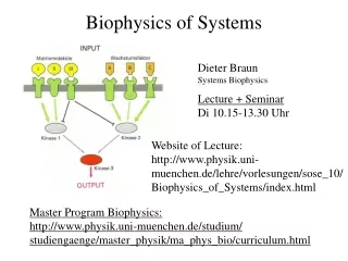
Biophysics Division Cellular/Molecular Imaging for Bonghan System Study 2006. 9. 27. Kwang-Sup Soh Biomedical Physics Lab. for Korean Medicine School of Physics, Seoul National University
Contents I. Introduction on Bonghan System II. Co-operative Research Progress Electron Microscope Atomic Force Microscope Nanoparticles
Superficial BH duct (Acupuncture meridian) Superficial BH Corpuscle (Acupoints) Organ-Surface BH duct (on the surface of organs) Bonghan System Intravascular BH duct skin : superficial BH ducts network surface of organs : OSBHD inside vessels : intra-vascular BHD
Physiological Significance • New Circulation System • Hyaluronic acid, Amino acid, Mono nucleotide, • Hormone: Adrenalin/noradrenalin • Bonghan Granules (Ф≈0.8~2.4μm) containing DNA • Adult stem cells: cell therapy • Natural “micro-cell”: gene therapy
Future Prospects Anatomy Physiology Electron Microscopes AFM Nanobiotechnology Optical Tweezer 2005 Sept, CKC Symposium
Electron Microscopes Microscopy Research & Technology, 2006 Electron Microscopic Study of Novel Threadlike Structures on the Surfaces of Mammalian Organs Byung-Cheon Lee,1 Jung Sun Yoo,1 Vyacheslav Ogay,1 Ki Woo Kim, 2Harald Dobberstein, 3 Kwang-Sup Soh, 1, † and Byung-Soo Chang 4, † 1Biomedical Physics Laboratory, FPRD, School of Physics and Astronomy, Seoul National University, Seoul, Korea2National Instrumentation Center for Environmental Management, College of Agriculture and Life Sciences, Seoul National University, Seoul, Korea3Department of Physics, Cavendish Laboratory, University of Cambridge, Cambridge, CB3 0HE, UK4Department of Cosmetology, Hanseo University, Seosan, Korea
Electron Microscopes Fig.1. Stereomicroscopic Images of novel threadlike structures
Electron Microscopes Cryo-SEM FIB/SEM (Cavendish)
Atomic Force Microscope Microcell-like granules in the Bonghan corpuscle on the surfaces of mammalian internal organs 1Ku Youn Baik, 1Vyacheslav Ogay, 2Harald Dobberstein, and 1Kwang-Sup Soh 1Biomedical Physics Laboratory, Department of Physics, Seoul National University, Seoul, 151-747, Korea 2Cavendish Laboratory, University of Cambridge, Cambridge, CB3 0HE, UK Investigation of morphology and physical properties of Bonghan microcell using AFM and TEM 1Ku Youn Baik, 1Joonhyung Kwon, 1Byung –Cheon Lee, 2Harald Dobberstein, and 1Kwang-Sup Soh$1Biomedical Physics Laboratory, Department of Physics, Seoul National University, Seoul, 151-747, Korea2Cavendish Laboratory, University of Cambridge, Cambridge, CB3 0HE, UK - 2006.7.2-7 Molecular Cell Biology GRC, NH, USA - 2006.8.27 WC2006 Conference, Seoul, Korea
Atomic Force Microscope Theory of Bonghan Microcell Bonghan microcell cycle (Bonghan Kim, 1965) (1) All the morphological components of organism are ceaselessly reproduced. (2) The self-reproduction of organism takes the form of Bonghan microcell-cell cycles. (3) The self-reproduction of organism is performed by the acupuncture-meridian system.
AFM image of Bonghan microcell A Topography, B Error, C Magnified of B, D line profile 1.4 um x 1um x 350nm sized oval Bonghan microcell. It shows clearly unique surface structure of layers and portions (B & C) whose size is from 0.2um to 0.6 um in diameter. The line profile analysis shows that the common patterns on the surface are steps of about 20nm height. This pattern is thought to be revealing the stereotype of its membrane structure. B A C D 20nm
Nanobiotechnology Flowing Channel of Nanoparticles