DNA molecule
Gene 2. DNA molecule. Gene 1. Gene 3. DNA template strand. TRANSCRIPTION. mRNA. Codon. TRANSLATION. Protein. Amino acid. Second mRNA base. First mRNA base (5 end of codon). Third mRNA base (3 end of codon). Wild-type. DNA template strand. 3 . 5 . 3 . 5 . mRNA. 5 .

DNA molecule
E N D
Presentation Transcript
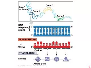
Gene 2 DNA molecule Gene 1 Gene 3 DNA template strand TRANSCRIPTION mRNA Codon TRANSLATION Protein Amino acid
Second mRNA base First mRNA base (5 end of codon) Third mRNA base (3 end of codon)
Wild-type DNA template strand 3 5 3 5 mRNA 5 3 Protein Stop Amino end Carboxyl end A instead of G Extra A 5 3 5 3 5 5 3 3 U instead of C Extra U 5 5 3 3 Stop Stop Silent (no effect on amino acid sequence) Frameshift causing immediate nonsense (1 base-pair insertion) T instead of C missing 3 3 5 5 5 5 3 3 A instead of G missing 5 3 5 3 Stop Missense Frameshift causing extensive missense (1 base-pair deletion) missing A instead of T 5 3 5 3 5 3 5 3 U instead of A missing 5 3 5 3 Stop Stop Nonsense No frameshift, but one amino acid missing (3 base-pair deletion) (a) Base-pair substitution (b) Base-pair insertion or deletion
Wild-type hemoglobin DNA Mutant hemoglobin DNA C T T C 3 3 A T 5 5 T 5 A G G A A 3 5 3 mRNA mRNA G A A 5 G U A 3 5 3 Normal hemoglobin Sickle-cell hemoglobin Val Glu
Normal hemoglobin Sickle-cell hemoglobin Primary structure His Val Leu Glu Glu His Thr Val Primary structure Thr Pro Val Leu Pro Glu 1 2 3 4 5 6 7 1 2 3 4 5 6 7 Exposed hydrophobic region Secondary and tertiary structures Secondary and tertiary structures subunit subunit Sickle-cell hemoglobin Quaternary structure Normal hemoglobin (top view) Quaternary structure Function Molecules interact with one another and crystallize into a fiber; capacity to carry oxygen is greatly reduced. Function Molecules do not associate with one another; each carries oxygen. 10 µm 10 µm Red blood cell shape Normal red blood cells are full of individual hemoglobin moledules, each carrying oxygen. Red blood cell shape Fibers of abnormal hemoglobin deform red blood cell into sickle shape.