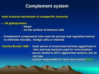
Complement
Complement. S. Barbour. Suggested Reading: Janeway, Chapter 2, pp. 61-82 Chapter 9, pp. 406-412. Office hours by arrangement: Please contact me by email (sbarbour@vcu.edu) to schedule an appointment. I. Overview of the Complement System. Hallmarks of Complement. Sequential Activation


Complement
E N D
Presentation Transcript
Complement S. Barbour Suggested Reading: Janeway, Chapter 2, pp. 61-82 Chapter 9, pp. 406-412 Office hours by arrangement: Please contact me by email (sbarbour@vcu.edu) to schedule an appointment.
Hallmarks of Complement • Sequential Activation • Amplification • Regulation
Overview of Complement Classical Pathway Alternative Pathway Lectin Pathway Antibody binds to specific antigen on pathogen surface Clearance of Apoptotic Cells Complement Activation Formation of C3 and C5 convertases Inflammatory response Mannose-binding protein binds pathogen surface Pathogen surface creates environment conducive to complement activation Opsonization & phagocytosis of some pathogens Clearance of Immune complexes Membrane Attack Pathway Cytolysis of some pathogens
Complement Nomenclature 1. CX (X = 1-9); example: C5 2. Factor X (alternative pathway); example: Factor B (B) Note: “i” denotes “inactive” fragments that do not support complement activation. However, these fragments can have other biological functions. (eg. iC3b or C3bi) 3. complement fragments (usually a and b); example: C3 → C3a + C3b Note: the “b” fragment remains cell associated; “a” fragment is soluble
C3 convertase C3 convertase C1 C1 C3b C3b C3 C3 C3b C3b Activation of the Classical Pathway IgG (IgG1, IgG2, IgG3) IgM Y Y Y Y Y Y Y Antigen (bacterial or yeast cell surface) -Note: only antigen-antibodies complexes can activate the classical pathway! Antibody alone cannot -Classical pathway results in formation of a C3 convertase (C4b2b) that generates C3b -C3b is covalently attached to pathogen; triggers complement activation and biological activity
C3 convertase C3b C3 C3b Activation of the Lectin Pathway mannan binding lectin (MBL) mannose sugars MBL “activating surface” (bacterial or yeast cell surface) -mannan binding lectin (MBL) recognizes mannose sugars on microbial cells -host mannose is hidden and is not accessible to MBL -lectin pathway results in the formation of a C3 convertase (C4b2b) that generates C3b
C3 convertase Soluble C3 convertase C1 C3b C3b C3 C3 C3b C3b Activation of the Alternative Pathway(Sources of C3b) C3 tickover Y Y classical pathway “activating surface” (bacterial or yeast cell surface) -the alternative pathway is initiated by C3b binding to a bacterial or yeast cell surface -this C3b can come from the classical pathway or “C3 tickover” -C3b preferentially interacts with bacterial or yeast cells; host cells are spared -this is a primitive distinction of self versus non-self
C3 convertase C3 convertase Soluble C3 convertase C3b C3b C3 C3 C3b C3b Activation of the Alternative Pathway(C3 Convertase) C3 tickover “activating surface” (bacterial or yeast cell surface) -alternative pathway results in the formation of a C3 convertase (C3bBb) that generates C3b -this is the same C3b that is produced by the classical pathway; two C3b molecules in alternative pathway -C3 tickover occurs constantly; regulatory proteins prevent C3b from binding host cells
C3 Convertase and C5 Convertase • C3 convertases generate C3b • C3b forms covalent attachment with pathogen surface • Together with other components, attached C3b forms C5 convertase • C5 convertase proteolyzes (activates) C5; last step in complement activation
Importance of C3 • Activation of Classical, Lectin, and Alternative pathways (alternative pathway is constantly activated in serum!) • Fragments have biological activity • Opsonization / Phagocytosis • B cell activation • Inflammation • Most abundant complement protein in serum ( > 1 mg/ml)
C3 C3a + C3b C2b C4b Bb C3b C3 Convertases C3 convertase Classical / Lectin Pathway Alternative Pathway receptor subunit receptor subunit catalytic subunit catalytic subunit P covalent association with pathogen surface -C3 convertases consist of two subunits; one with catalytic activity, one that binds C3 -subunits must be associated to have active C3 convertase -regulatory mechanisms are designed to disrupt this association
Regulation of C3 Convertases Dissociation Proteolysis (Factor I cofactor) In general, regulatory proteins are expressed on host cells, but not on bacteria. Therefore, host cells are spared from complement attack.
The Membrane Attack Pathway MAC The MAC is especially important for the immune response against Neisseria spp. Membrane proteins (CD59, HRF) prevent MAC formation on host cells.
opsonized bacterium Opsonization / Phagocytosis -both C3b and iC3b (fragment of C3b) are opsonins -in addition to CR1, CR3 and CR4 also mediate phagocytosis
Inflammation anaphylotoxins C3a, C4a---increased vascular permeability C5a—chemoattraction C3a, C4a----activation
Complement Proteins and Inflammation • C4a, C3a, and C5a are pro-inflammatory “anaphylotoxins” • C5a binds a G protein coupled receptor • -neutrophils express high levels of the C5a receptor • -C5a binding induces chemotaxis and activation / • increased cytocidal activity • C3a, C4a bind another G protein coupled receptor • -C3a/4a receptor is expressed by phagocytes, endothelial • cells • -C3a/ C4a binding induces cell activation, increased • cytocidal activity, increased vascular permeability
B cell Activation or C3dg -microbial cell expresses antigens recognized by B cell receptor -microbial cell is coated with C3d or C3dg (fragments of C3b) -C3d/ C3dg is recognized by CR2 -simultaneous binding to B cell receptor and CR2 results in efficient B cell activation
Clearance of Apoptotic Cells C1qR C1q phagocyte • Phagocyte recognizes C1q deposited on the surface of apoptotic cell • Apoptotic cell is ingested and destroyed by phagocyte • This may be an important mechanism for clearing self antigens -Alternatively, uptake of apoptotic host cells by C1q on immature DC may induce self tolerance - prevalence of autoimmune disease is very high in subjects lacking C1q
Classical Pathway Alternative Pathway Lectin Pathway Antibody binds to specific antigen on pathogen surface Clearance of Apoptotic Cells Complement Activation Formation of C3 and C5 convertases Inflammatory response Mannose-binding protein binds pathogen surface Membrane Attack Pathway Cytolysis of some pathogens Pathogen surface creates environment conducive to complement activation Opsonization & phagocytosis Of some pathogens Clearance of Immune complexes Overview of Complement
Clearance of apoptotic cells C3d C3dg B cell activation