Download
1 / 84
930 likes | 1.4k Vues
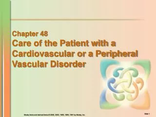
Chapter 48 Care of the Patient with a Cardiovascular or a Peripheral Vascular Disorder. Overview ofAnatomy and Physiology. Heart Four-chambered, hollow, muscular organ, not much bigger than a fist Lies in the mediastinum Lower border is called the apex Heart wall: three layers

E N D
Chapter 48 Care of the Patient with a Cardiovascular or a Peripheral Vascular Disorder
Overview ofAnatomy and Physiology • Heart • Four-chambered, hollow, muscular organ, not much bigger than a fist • Lies in the mediastinum • Lower border is called the apex • Heart wall: three layers • Epicardium: serous membrane on the outside of the heart • Myocardium: constructed of cardiac muscle • Endocardium: lines the inner surface of the chambers of the heart
Figure 48-1 (From Thibodeau, G.A., Patton, K.T. [1987]. Anatomy and physiology. St. Louis: Mosby.) Heart and major blood vessels viewed from front (anterior).
Overview of Anatomy and Physiology • Heart chambers • Right atrium—receives deoxygenated blood • Left atrium—receivesoxygenated blood • Right ventricle—pumps deoxygenated blood • Left ventricle—pumps oxygenated blood • Heart valves • Atrioventricular valves ( AV) valves • Tricuspid and bicuspid valves • Semilunar valves • Pulmonary and aortic semilunar valves
Figure 48-2 (From Thibodeau, G.A., Patton, K.T. [1987]. Anatomy and physiology. St. Louis: Mosby.) Interior of the heart.
Overview of Anatomy and PhysiologyNew Slide 1. Identify the four main types of tissues which make up the body’s major organs. 2. How does skeletal, smooth ( visceral)and cardiac muscle cells differ in ref: appearance, movement and location?
Electrical conduction system • Automaticity • An inherent ability of the heart muscle tissue to contract in a rhythmic pattern • Irritability • The ability to respond to a stimulus • Impulse pattern • Sinoatrial node to AV node to bundle of His to right and left bundle branches to Purkinje fibers
Figure 48-3 (new) Located w/in the walls of the heart & septum Identify the Conduction system structures 1. 2. 3. 4. 5. (From Thibodeau, G.A., Patton, K.T. [1987]. Anatomy and physiology. St. Louis: Mosby.) Conduction system of the heart.
Overview of Anatomy and Physiology • Cardiac cycle • A complete heartbeat • Atria contract while ventricles relax • Ventricles contract while atria relax • Systole • Phase of contraction • Diastole • Phase of relaxation • Period between contraction of the atria or ventricles during which the blood enters the relaxed chambers • A complete cycle takes an average of 0.8 second
Figure 48-4 (From Canobbio, M. [1990]. Cardiovascular disorders, Mosby’s clinical nursing series. St. Louis: Mosby.) Blood flow during systole.
Figure 48-5 (From Canobbio, M. [1990]. Cardiovascular disorders, Mosby’s clinical nursing series. St. Louis: Mosby.) Blood flow during diastole.
Review Electrocardiography Figure 48-7 True or False Atrial repolarization is captured on the EKG? Explain. So what causes cardiac impulse to occur? Ch. 15 & ch. 10A&P book Normal ECG deflections.
Overview of Anatomy and Physiology • Blood vessels ( 3 main types) • Capillaries • Tiny blood vessels joining arterioles and venules • How are capillaries able to produce gas exchange? • Arteries • Large vessels carrying blood away from the heart • Veins • Vessels that convey blood from the capillaries to the heart
Circulation • Coronary blood supply • Right and left coronary arteries • Branch off of the aorta • Encircle the heart like a crown • Supply the myocardium with blood • Coronary veins • Return the unoxygenated blood to the coronary sinus, then to the right atrium • When might collateral circulation become necessary?
Figure 48-6 (From Canobbio, M. [1990]. Cardiovascular disorders, Mosby’s clinical nursing series. St. Louis: Mosby.) Arterial coronary circulation (anterior).
Circulation • Systemic circulation • Circulates blood from the left ventricle to all parts of the body and back to the right atrium • Carries oxygen and nutritive materials to all body tissues and removes products of metabolism • Pulmonary circulation • Circulates blood from the right ventricle to the lungs and back to the left atrium of the heart • Carries deoxygenated blood to the lungs to be reoxygenated and removes the metabolic waste product, carbon dioxide
Laboratory and Diagnostic Examinations • Diagnostic imaging • Fluoroscopy • Angiogram • Aortogram • Cardiac catheterization and angiography • Electrocardiography • Cardiac monitors • Thallium scanning • Laboratory tests: CBC, blood cultures, coagulation studies, ESR electrolytes, lipids, arterial blood gases, cardiac markers
Disorders of the Cardiovascular System • Risk factors • Non-modifiable factors • Family history • Age • Sex (gender) • Race
Disorders of the Cardiovascular System • Risk factors (continued) • Modifiable factors • Smoking • Hyperlipidemia • Hypertension • Diabetes mellitus • Obesity • Sedentary lifestyle • Stress • Oral contraceptives • Psychosocial factors
Disorders of the Cardiovascular System • Cardiac dysrhythmias • Any cardiac rhythm that deviates from normal sinus rhythm • Sinus tachycardia • Sinus bradycardia • Supraventricular tachycardia • Atrial fibrillation • Atrioventricular block • Premature ventricular contractions • Ventricular tachycardia • Ventricular fibrillation
Disorders of the Cardiovascular System • Cardiac Arrest • The sudden cessation of cardiac output and circulatory process • Cause: ventricular tachycardia, ventricular fibrillation, and ventricular asystole • S/S: abrupt loss of consciousness with no response to stimuli; gasping respirations followed by apnea; absence of pulse and blood pressure; pupil dilation; pallor and cyanosis • Tx: • Immediate CPR ( CAB) no longer ABCs • ACLS • Review Medications for Cardiac Dysrhythmias
Disorders of the Heart • Coronary atherosclerotic heart disease • Coronary artery disease (CAD) • A variety of conditions that obstruct blood flow in the coronary arteries • Atherosclerosis • A common arterial disorder characterized by yellowish plaques of cholesterol, lipids, and cellular debris in the inner layers of the walls of the arteries; the primary cause of atherosclerotic heart disease (ASHD)
Figure 48-10 (From Lewis, S.M., Heitkemper, M.M., Dirksen, S.R. [2004]. Medical-surgical nursing: assessment and management of clinical problems. [6th ed.]. St. Louis: Mosby.) Progressive development of coronary atherosclerosis.
Disorders of the Heart • Angina pectoris • Etiology/pathophysiology • Cardiac muscle is deprived of oxygen • Increased workload on the heart • Clinical manifestations/assessment • Pain (usually relieved by rest) • Dyspnea • Anxiety; apprehension • Diaphoresis • Nausea
Disorders of the Heart • Angina pectoris (continued) • Medical management/nursing interventions • Correct cardiovascular risk factors • Avoid precipitating factors • Medications • Dilate coronary arteries and decrease workload of heart • Nitroglycerin • Beta-adrenergic blocking agents • Calcium channel blockers
Disorders of the Heart • Angina pectoris (continued) • Medical management/nursing interventions • Surgical interventions • Coronary artery bypass graft (CABG) • Percutaneous transluminal coronary angioplasty (PTCA) • Stent placement
Disorders of the Heart • Myocardial infarction • Etiology/pathophysiology • Occlusion of a major coronary artery or one of its branches with subsequent necrosis of myocardium • Most common cause is atherosclerosis • Ability of the cardiac muscle to contract and pump blood is impaired
Figure 48-16 (From Lewis, S.M., Heitkemper, M.M., Dirksen, S.R. [2004]. Medical-surgical nursing: assessment and management of clinical problems. [6th ed.]. St. Louis: Mosby.) Four common locations where myocardial infarctions occur.
Disorders of the Heart • Myocardial infarction (continued) • Clinical manifestations/assessment • Asymptomatic (silent MI) • Pain (not relieved by rest, position, or nitroglycerin) • Nausea • SOB; dizziness; weakness • Diaphoresis • Pallor—ashen color • Sense of impending doom
Figure 48-11 (From Lewis, S.M., Heitkemper, M.M., Dirksen, S.R. [2004]. Medical-surgical nursing: assessment and management of clinical problems. [6th ed.]. St. Louis: Mosby.) Sites to which ischemic myocardial pain may be referred.
Disorders of the Heart • Myocardial infarction (continued) • Medical management/nursing interventions • Oxygen • Fibrinolytic agents • Percutaneous transluminal coronary angioplasty (PTCA) • Coronary artery bypass graft surgery • Medications: vasopressors, analgesics, nitrates, beta-adrenergic blockers, calcium channel blockers, antidysrhythmics, diuretics, inotropic agents, diuretics, stool softeners
Figure 48-12 (A, from Thelan, L., et al. [1998]. Critical care nursing. [3rd ed.]. St. Louis: Mosby. B, from Lewis, S.M., Heitkemper, M.M., Dirksen, S.R. [2004]. Medical-surgical nursing: assessment and management of clinical problems. [6th ed.]. St. Louis: Mosby.) A, Saphenous vein. B, Saphenous aortocoronary artery bypass.
Figure 48-13 (From Phipps, W., et al. [1995]. Medical-surgical nursing: concepts and clinical practice. [5th ed.]. St. Louis: Mosby.) Coronary artery bypass graft.
Disorders of the Heart • Heart failure • Etiology/pathophysiology • Abnormal condition characterized by circulatory congestion resulting from the heart’s inability to act as an effective pump • Left ventricular failure • Most common • Right ventricular failure • Usually caused by left ventricular failure
Disorders of the Heart • Heart failure (continued) • Clinical manifestations/assessment • Decreased cardiac output • Fatigue • Angina • Anxiety; restlessness • Oliguria • Decreased GI motility • Pale, cool skin • Weight gain
Disorders of the Heart • Heart failure (continued) • Clinical manifestations/assessment (continued) • Left ventricular failure • Pulmonary congestion • Dyspnea • Paroxysmal nocturnal dyspnea • Cough; frothy, blood-tinged sputum • Orthopnea • Pulmonary crackles • Pleural effusion (x-ray)
Disorders of the Heart • Heart failure (continued) • Clinical manifestations/assessment (continued) • Right ventricular failure • Distended jugular veins • Anorexia, nausea, and abdominal distention • Liver enlargement • Ascites • Edema in feet, ankles, sacrum; may progress up the legs into thighs, external genitalia, and lower trunk
Disorders of the Heart • Heart failure (continued) • Medical management/nursing interventions • Increase cardiac efficiency • Digitalis • Vasodilators • ACE inhibitors (decrease blood pressure) • Bedrest, HOB elevated • Oxygen • Treat edema and pulmonary congestion • Monitor fluid retention (weigh daily; strict I&O)
Disorders of the Heart • Pulmonary edema • Etiology/pathophysiology • Accumulation of fluid in lung tissues and alveoli • Complication of congestive heart failure (CHF) • Clinical manifestations/assessment • Restlessness • Agitation • Disorientation • Diaphoresis • Dyspnea and tachypnea
Disorders of the Heart • Pulmonary edema (continued) • Clinical manifestations/assessment (continued) • Tachycardia • Pallor or cyanosis • Cough—large amounts of blood-tinged, frothy sputum • Wheezing, crackles • Cold extremities
Disorders of the Heart • Pulmonary edema (continued) • Medical management/nursing interventions • High Fowler’s or orthopnic position • Morphine sulfate • Oxygen • Nitroglycerin • Diuretics • Inotropic agents • Vasodilators
Disorders of the Heart • Valvular heart disease • Etiology/pathophysiology • Heart valves are compromised and do not open and close properly • Stenosis • Insufficiency • Causes may be: • Congenital • Rheumatic fever
Disorders of the Heart • Valvular heart disease (continued) • Clinical manifestations/assessment • Fatigue • Angina • Oliguria • Pale, cool skin • Weight gain • Restlessness • Abnormal breath sounds • Edema
Disorders of the Heart • Valvular heart disease (continued) • Medical management/nursing interventions • Restrict activities • Sodium-restricted diet • Diuretics • Digoxin • Antidysrhythmics • Surgery • Open mitral commissurotomy • Valve replacement
Disorders of the Heart • Rheumatic heart disease • Etiology/pathophysiology • Rheumatic fever • Inflammatory disease which is a delayed childhood reaction to inadequately treated childhood upper respiratory tract infection of beta-hemolytic streptococci • Causes scar tissue in the heart
Disorders of the Heart • Rheumatic heart disease (continued) • Clinical manifestations/assessment • Elevated temperature • Elevated heart rate • Epistaxis • Anemia • Joint pain and stiffness • Nodules on the joints • Specific to valve affected • Heart murmur
Disorders of the Heart • Rheumatic heart disease (continued) • Medical management/nursing interventions • Prevention • Treat infections rapidly and completely • Bedrest • NSAIDs • Application of heat • Well-balanced diet (supplement with vitamins B and C) • Encourage fluids • Commissurotomy or valve replacement
Disorders of the Heart • Pericarditis • Etiology/pathophysiology • Inflammation of the membranous sac surrounding the heart • May be acute or chronic • Bacterial, viral, or fungal • Noninfectious conditions • Azotemia, MI, neoplasms, scleroderma, trauma, systemic lupus erythematosus (SLE), radiation, drugs
Disorders of the Heart • Pericarditis (continued) • Clinical manifestations/assessment • Debilitating pain • Dyspnea • Fever • Chills • Diaphoresis • Leukocytosis • Pericardial friction rub • Pericardial effusion
Disorders of the Heart • Pericarditis (continued) • Medical management/nursing interventions • Analgesia • Oxygen • IV fluids • Salicylates • Antibiotics • Antiinflammatory agents • Corticosteroids • Surgery: pericardial window, pericardial tap