CELL COMMUNICATION
CELL COMMUNICATION. Section A: An Overview of Cell Signaling. Section A: An Overview of Cell Signaling. 1. Cell signaling evolved early in the history of life 2. Communicating cells may be close together or far apart

CELL COMMUNICATION
E N D
Presentation Transcript
CELL COMMUNICATION Section A: An Overview of Cell Signaling Lecture 8 - Chapter 11
Section A: An Overview of Cell Signaling • 1. Cell signaling evolved early in the history of life • 2. Communicating cells may be close together or far apart • 3. The three stages of cell signaling are reception, transduction, and response Lecture 8 - Chapter 11
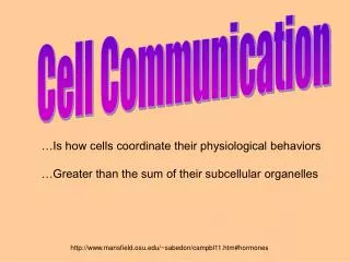
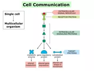
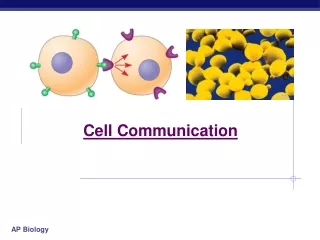
Introduction • Cell-to-cell communication is absolutely essential for multicellular organisms. • Cells must communicate to coordinate their activities. • Communication between cells is also important for many unicellular organisms. • Biologists have discovered some universal mechanisms of cellular regulation, involving the same small set of cell-signaling mechanisms. • Cells may receive a variety of signals, chemical signals, electromagnetic signals, and mechanical signals. Lecture 8 - Chapter 11
1. Cell signaling evolved early in the history of life • One topic of cell “conversation” is sex. • The yeast Saccharomycescerevisiae, the yeast of bread, wine, and beer, identifies its mates by chemical signaling. • There are two sexes, a and alpha, each of which secretes a specific signaling molecule, a factor and alpha factor respectively. • These factors each bind to receptor proteins on the other mating type. Lecture 8 - Chapter 11
Once the mating factors have bound to the receptors, the two cells grow toward each other and experience other cellular changes. • Two opposite cells fuse, or mate. • The a/alpha cell contains the genes of both cells. Lecture 8 - Chapter 11
The process by which a signal on a cell’s surface is converted into a specific cellular response consists of a series of steps called a signal-transduction pathway. • The molecular details in both yeast and animal cells are strikingly similar, even though their last common ancestor was over a billion years ago. • Signaling molecules evolved first in ancient prokaryotes and were then adopted for new uses by single-celled eukaryotes and multicellular descendents. Lecture 8 - Chapter 11
Cell signaling has remained important in the microbial world. • Myxobacteria, soil-dwelling bacteria, use chemical signals to communicate nutrient availability. • When food is scarce, cells secrete a signal to other cells leading them to aggregate and form thick-walled spores. Lecture 8 - Chapter 11
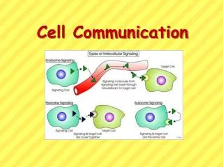
2. Communicating cells may be close together or far apart • Multicellular organisms also release signaling molecules that target other cells. • Some transmitting cells release local regulators that influence cells in the local vicinity. • Paracrine signaling occurs when numerous cells can simultaneously receive and respond to growth factors produced by a single cell in their vicinity. Lecture 8 - Chapter 11
In synaptic signaling, a nerve cell produces a neurotransmitter that diffuses to a single cell that is almost touching the sender. • An electrical signal passing along the nerve cell triggers secretion of the neurotransmitter into the synapse. • Nerve signals can travel along a series of nerve cells without unwanted responses from other cells. Lecture 8 - Chapter 11
Plants and animals use hormones to signal at greater distances. • In animals, specialized endocrine cells release hormones into the circulatory system, by which they travel to target cells in other parts of the body. • In plants, hormones may travel in vessels, but more often travel from cell to cell or by diffusion in air. Lecture 8 - Chapter 11
Hormones and local regulators range widely in size and type. • The plant hormone ethylene (C2H4), which promotes fruit ripening and regulates growth, is a hydrocarbon with only six atoms. • Insulin, which regulates sugar levels in the blood of mammals, is a protein with thousands of atoms. Lecture 8 - Chapter 11
Cells may communicate by direct contact. • Signaling substances dissolved in the cytosol pass freely between adjacent cells. • Cells may also communicate via direct contact between substances on their surfaces. Lecture 8 - Chapter 11
3. The three stages of cell signaling are reception, transduction, and response • The origins of our understanding of cell signaling were pioneered by E.W. Sutherland and his colleagues. • Their work investigated how the animal hormone epinephrine stimulates breakdown of the storage polysaccharide glycogen in liver and skeletal muscle. • Breakdown of glycogen releases glucose derivatives that can be used for fuel in glycolysis or released as glucose in the blood for fuel elsewhere. • One effect of the release of epinephrine from the adrenal gland is mobilization of fuel reserves. Lecture 8 - Chapter 11
Sutherland’s research team discovered that epinephrine activated a cytosolic enzyme, glycogen phosphorylase. • However, epinephrine did not activate the phosphorylase directly but could only act onintact cells. • Therefore, there must be an intermediate step or steps occurring inside the cell. • Also, the plasma membrane must be involved in transmitting the epinephrine signal. Lecture 8 - Chapter 11
The process must involve three stages. • In reception, a chemical signal binds to a cellular protein, typically at the cell’s surface. • In transduction, binding leads to a change in the receptor that triggers a series of changes along a signal-transduction pathway. • In response, the transduced signal triggers a specific cellular activity. Lecture 8 - Chapter 11
CELL COMMUNICATION Section B: Signal Reception and the Initiation of Transduction Lecture 8 - Chapter 11
Section B: Signal Reception and the Initiation of Transduction • 1. A signal molecule binds to a receptor protein, causing the protein to change shape • 2. Most signal receptors are plasma membrane proteins Lecture 8 - Chapter 11
1. A signal molecule binds to a receptor protein causing the protein to change shape • A cell targeted by a particular chemical signal has a receptor protein that recognizes the signal molecule. • Recognition occurs when the signal binds to a specific site on the receptor because it is complementary in shape. • When ligands (small molecules that bind specifically to a larger molecule) attach to the receptor protein, the receptor typically undergoes a change in shape. • This may activate the receptor so that it can interact with other molecules. • For other receptors, this leads to the aggregation of receptors. Lecture 8 - Chapter 11
2. Most signal receptors are plasma membrane proteins • Most signal molecules are water-soluble and too large to pass through the plasma membrane. • They influence cell activities by binding to receptor proteins on the plasma membrane. • Binding leads to change in the shape of the receptor or to the aggregation of receptors. • These trigger changes in the intracellular environment. • Three major types of receptors are G-protein-linked receptors, tyrosine-kinase receptors, and ion-channel receptors. Lecture 8 - Chapter 11
A G-protein-linked receptor consists of a receptor protein associated with a G-protein on the cytoplasmic side. • The receptor consists of seven alpha helices spanning the membrane. • Effective signal molecules include yeast mating factors, epinephrine, other hormones, and neurotransmitters. Lecture 8 - Chapter 11
The G protein acts as an on-off switch. • If GDP is bound, the G protein is inactive. • If ATP is bound, the G protein is active. Lecture 8 - Chapter 11
The G-protein system cycles between on and off. • When a G-protein-linked receptor is activated by binding with an extracellular signal molecule, the receptor binds to an inactive G protein in membrane. • This leads the G protein to substitute GTP for GDP. • The G protein then binds with another membrane protein, often an enzyme, altering its activity and leading to a cellular response. Lecture 8 - Chapter 11
The G protein can also act as a GTPase enzyme and hydrolyzes the GTP, which activated it, to GDP. • This change turns the G protein off. • The whole system can be shut down quickly when the extracellular signal molecule is no longer present. Lecture 8 - Chapter 11
G-protein receptor systems are extremely widespread and diverse in their functions. • In addition to functions already mentioned, they play an important role during embryonic development and sensory systems. • Similarities among G proteins and G-protein-linked receptors suggest that this signaling system evolved very early. • Several human diseases are the results of activities, including bacterial infections, which interfere with G-protein function. Lecture 8 - Chapter 11
The tyrosine-kinase receptor system is especially effective when the cell needs to regulate and coordinate a variety of activities and trigger several signal pathways at once. • Extracellular growth factors often bind to tyrosine-kinase receptors. • The cytoplasmic side of these receptors function as a tyrosine kinase, transferring a phosphate group from ATP to tyrosine on a substrate protein. Lecture 8 - Chapter 11
An individual tyrosine-kinase receptor consists of several parts: • an extracellular signal-binding sites, • a single alpha helix spanning the membrane, and • an intracellular tail with several tyrosines. Lecture 8 - Chapter 11
When ligands bind to two receptor polypeptides, the polypeptides aggregate, forming a dimer. • This activates the tyrosine-kinase section of both. • These add phosphates to the tyrosine tails of the other polypeptide. Lecture 8 - Chapter 11
The fully-activated receptor proteins activate a variety of specific relay proteins that bind to specific phosphorylated tyrosine molecules. • One tyrosine-kinase receptor dimer may activate ten or more different intracellular proteins simultaneously. • These activated relay proteins trigger many different transduction pathways and responses. Lecture 8 - Chapter 11
Ligand-gated ion channels are protein pores that open or close in response to a chemical signal. • This allows or blocks ion flow, such as Na+ or Ca2+. • Binding by a ligand to the extracellular side changes the protein’s shape and opens the channel. • Ion flow changes the concentration inside the cell. • When the ligand dissociates, the channel closes. Lecture 8 - Chapter 11
Ligand-gated ion channels are very important in the nervous system. • Similar gated ion channels respond to electrical signals. Lecture 8 - Chapter 11
Other signal receptors are dissolved in the cytosol or nucleus of target cells. • The signals pass through the plasma membrane. • These chemical messengers include the hydrophobic steroid and thyroid hormones of animals. • Also in this group is nitric oxide (NO), a gas whose small size allows it to slide between membrane phospholipids. Lecture 8 - Chapter 11
Testosterone, like other hormones, travels through the blood and enters cells throughout the body. • In the cytosol, they bind and activate receptor proteins. • These activated proteins enter the nucleus and turn on genes that control male sex characteristics. Lecture 8 - Chapter 11
These activated proteins act as transcription factors. • Transcription factors control which genes are turned on - that is, which genes are transcribed into messenger RNA (mRNA). • The mRNA molecules leave the nucleus and carry information that directs the synthesis (translation) of specific proteins at the ribosome. • Other intracellular receptors are already in the nucleus and bind to the signal molecules there (e.g., estrogen receptors). Lecture 8 - Chapter 11
CELL COMMUNICATION Section C: Signal-Transduction Pathways Lecture 8 - Chapter 11
Section C: Signal-Transduction Pathways • 1. Pathways relay signals from receptors to cellular responses • 2. Protein phosphorylation, a common mode of regulation in cells, is a major mechanism of signal transduction • 3. Certain small molecules and ions are key components of signaling pathways (second messengers) Lecture 8 - Chapter 11
Introduction • The transduction stage of signaling is usually a multistep pathway. • These pathways often greatly amplify the signal. • If some molecules in a pathway transmit a signal to multiple molecules of the next component, the result can be large numbers of activated molecules at the end of the pathway. • A small number of signal molecules can produce a large cellular response. • Also, multistep pathways provide more opportunities for coordination and regulation than do simpler systems. Lecture 8 - Chapter 11
1. Pathways relay signals from receptors to cellular responses • Signal-transduction pathways act like falling dominoes. • The signal-activated receptor activates another protein, which activates another and so on, until the protein that produces the final cellular response is activated. • The original signal molecule is not passed along the pathway and may not even enter the cell. • Its information is passed on. • At each step the signal is transduced into a different form, often by a conformational change in a protein. Lecture 8 - Chapter 11
2. Protein phosphorylation, a common mode of regulation in cells, is a major mechanism of signal transduction • The phosphorylation of proteins by a specific enzyme (a protein kinase) is a widespread cellular mechanism for regulating protein activity. • Most protein kinases act on other substrate proteins, unlike the tyrosine kinases that act on themselves. • Most phosphorylation occurs at either serine or threonine amino acids of the substrate protein. Lecture 8 - Chapter 11
Many of the relay molecules in a signal-transduction pathway are protein kinases that lead to a “phosphorylation cascade”. • Each protein phosphorylation leads to a shape change because of the interaction between the phosphate group and charged or polar amino acids. Lecture 8 - Chapter 11
Phosphorylation of a protein typically converts it from an inactive form to an active form. • The reverse (inactivation) is possible too for some proteins. • A single cell may have hundreds of different protein kinases, each specific for a different substrate protein. • Fully 1% of our genes may code for protein kinases. • Abnormal activity of protein kinases can cause abnormal cell growth and contribute to the development of cancer. Lecture 8 - Chapter 11
The responsibility for turning off a signal-transduction pathway belongs to protein phosphatases. • These enzymes rapidly remove phosphate groups from proteins. • The activity of a protein regulated by phosphorylation depends on the balance of active kinase molecules and active phosphatase molecules. • When an extracellular signal molecule is absent, active phosphatase molecules predominate, and the signaling pathway and cellular response are shut down. Lecture 8 - Chapter 11
3. Certain signal molecules and ions are key components of signaling pathways (second messengers) • Many signaling pathways involve small, nonprotein, water-soluble molecules or ions, called second messengers. • These molecules rapidly diffuse throughout the cell. • Second messengers participate in pathways initiated by both G-protein-linked receptors and tyrosine-kinase receptors. • Two of the most important are cyclic AMP and Ca2+. Lecture 8 - Chapter 11
Once Sutherland knew that epinephrine caused glycogen breakdown without entering the cell, he looked for a second messenger inside the cell. • Binding by epinephrine leads to increases in the concentration of cyclic AMP or cAMP. • This occurs because the receptor activates adenylyl cyclase, which converts ATP to cAMP. • cAMP is short-lived as phosphodiesterase converts it to AMP. Lecture 8 - Chapter 11
More generally, many hormones and other signals trigger the formation of cAMP. • Binding by the signal to a receptor activates a G protein that activates adenylyl cyclase in the plasma membrane. • The cAMP from the adenylyl cyclase diffuses through the cell and activates a serine/threonine kinase, called protein kinaseAwhich phosphorylates other proteins. Lecture 8 - Chapter 11
Other G-protein systems inhibit adenylyl cyclase. • These use a different signal molecule to activate other receptors that activate inhibitory G proteins. • Certain microbes cause disease by disrupting the G-protein signaling pathways. • The cholera bacterium, Vibrio cholerae, colonizes the small intestine and produces a toxin that modifies a G protein that regulates salt and water secretion. • The modified G protein is stuck in its active form, continuously stimulating productions of cAMP. • This causes the intestinal cells to secrete large amounts of water and salts into the intestines, leading to profuse diarrhea and death if untreated. Lecture 8 - Chapter 11
Many signal molecules in animals induce responses in their target cells via signal-transduction pathways that increase the cytosolic concentration of Ca2+. • In animal cells, increases in Ca2+ may cause contraction of muscle cells, secretion of some substances, and cell division. • In plant cells, increases in Ca2+ trigger responses for coping with environmental stress, including drought. • Cells use Ca2+ as a second messenger in both G-protein pathways and tyrosine-kinase pathways. Lecture 8 - Chapter 11
The Ca2+ concentration in the cytosol is typically much lower than that outside the cell, often by a factor of 10,000 or more. • Various protein pumps transport Ca2+ outside the cell or inside the endoplasmic reticulum or other organelles. Lecture 8 - Chapter 11
Because cytosolic Ca2+ is so low, small changes in the absolute numbers of ions causes a relatively large percentage change in Ca2+ concentration. • Signal-transduction pathways trigger the release of Ca2+ from the cell’s ER. • The pathways leading to release involve still other second messengers, diacylglycerol (DAG) and inositol trisphosphate (IP3). • Both molecules are produced by cleavage of certain phospholipids in the plasma membrane. Lecture 8 - Chapter 11
DAG and IP3 are created when a phospholipase cleaves a membrane phospholipid PIP2. • Phospholipase may be activated by a G protein or by a tyrosine-kinase receptor. • IP3 activates a gated-calcium channel, releasing Ca2+. Lecture 8 - Chapter 11