Download
1 / 22
220 likes | 244 Vues
Case Study: Radiation Therapy and Ultrasound Management of Breast Cancer. hhholdorf. Radiation Therapy (also known as radiotherapy and radiation oncology) began shortly after the discovery of X-rays in 1895 by Wilhelm Rontgen.

E N D
Case Study:Radiation Therapy and Ultrasound Management of Breast Cancer hhholdorf
Radiation Therapy (also known as radiotherapy and radiation oncology) began shortly after the discovery of X-rays in 1895 by Wilhelm Rontgen. • In 1896, Antoine-Henri Becquerel discovered that certain elements spontaneously emitted rays or subatomic particles from matter, a property which came to be known as radioactivity. • Building on the work of Becquerel, Pierre and Marie Curie discovered the radioactive elements polonium and radium. While experimenting, they noticed that radium killed diseased cells -- the first indication that radiation could aid not just in the diagnosis of disease, but also in treatment. Background and History
Due to the groundbreaking work of Nobel Prize-winning scientists Antoine-Henri Becquerel, Marie Curie and Pierre Curie, the field of radiation therapy grew quickly in the early 1900s. A new era in medical treatment and research began. Background and History
What is radiation therapy? • Radiation therapy is a form of cancer treatment that uses radiation (strong beams of energy) to kill cancer cells or keep them from growing and dividing. Background and History
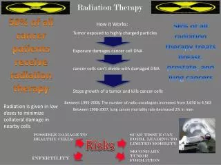
How does radiation therapy kill cancer cells? • Radiation therapy kills cancer cells by damaging their DNA (the molecules inside cells that carry genetic information and pass it from one generation to the next). Radiation therapy can either damage DNA directly or create charged particles (free radicals) within the cells that can in turn damage the DNA. • Cancer cells whose DNA is damaged beyond repair stop dividing or die. When the damaged cells die, they are broken down and eliminated by the body’s natural processes. Background and History
Does radiation therapy kill only cancer cells? • No, radiation therapy can also damage normal cells, leading to side effects. Doctors take potential damage to normal cells into account when planning a course of radiation therapy. The amount of radiation that normal tissue can safely receive is known for all parts of the body. Doctors use this information to help them decide where to aim radiation during treatment. Background and History
Why do patients receive radiation therapy? • Radiation therapy is sometimes given with curative intent (that is, with the hope that the treatment will cure a cancer, either by eliminating a tumor, preventing cancer recurrence, or both). In such cases, radiation therapy may be used alone or in combination with surgery, chemotherapy, or both. • Radiation therapy may also be given with palliative intent. Palliative treatments are not intended to cure. Instead, they relieve symptoms and reduce the suffering caused by cancer. Background and History
What are the potential side effects of radiation therapy? • Radiation therapy can cause both early (acute) and late (chronic) side effects. Acute side effects occur during treatment, and chronic side effects occur months or even years after treatment ends. The side effects that develop depend on the area of the body being treated, the dose given per day, the total dose given, the patient’s general medical condition, and other treatments given at the same time. Fatigue is a common side effect of radiation therapy regardless of which part of the body is treated. Nausea with or without vomiting is common when the abdomen is treated and occurs sometimes when the brain is treated. Medications are available to help prevent or treat nausea and vomiting during treatment. Background and History
What are the potential side effects of radiation therapy? Acute Side Effects • Skin irritation, or damage at regions exposed to the radiation beams. Acute radiation side effects are caused by damage to rapidly dividing normal cells in the area being treated. Most acute effects disappear after treatment ends, though some can be permanent. Late Side Effects • Fibrosis (the replacement of normal tissue with scar tissue, leading to restricted movement of the affected area). • Damage to the bowels, causing diarrhea and bleeding. • Memory loss. • Infertility (inability to have a child). • Rarely, a second cancer caused by radiation exposure. Late side effects of radiation therapy may or may not occur. Depends on the area of the body treated Background and History
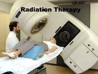
Radiation therapy of the breast Background and History
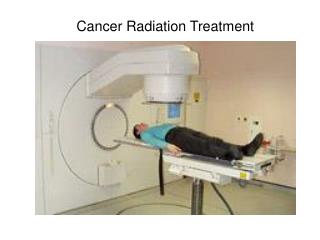
Radiation therapy of the pelvis. Lasers and a mold under the legs are used to determine exact position Background and History
Purpose • The processes was to create an individualized exercise program, and monitor training of a breast cancer survivor who was participating in a fitness plan during and after surgery, chemotherapy • Radiation treatments were examined over a 391-day period Case Study: Exercise capacity of a breast cancer survivor
Case Study • A 57-yr-old female was diagnosed with stage I breast cancer (approximately 1.2 cm diameter) with no lymph node involvement. After lumpectomy and axillary node dissection, the client completed chemotherapy treatment followed by 33 bouts of radiation therapy • Assessment included body composition, lactate threshold, VO2 max, pulmonary function testing which was measured 4 days post-diagnosis and 2 months after treatments had ended. The client kept a daily log of exercise, average heart rate, and rating of perceived exertion in each exercise session Case Study
Definitions • Lumpectomy- surgery in which only the tumor and some surrounding tissue is removed • Axillary node dissection- removal of nodes in levels depending on the case • VO2 Max- maximum capacity of an individuals body to transport and use oxygen during incremental exercise Case Study
Results • Over 391 calendar days, the client exercised 343 days and completed 424 exercise sessions. The client's body composition and body weight remained stable for the entire period. There was a significant decrease in VO2max before and after treatment. During the treatment phase, the client averaged 1.19 exercise sessions per day, with an average duration of 48 min at approximately 57% of VO2max. Post-treatment, the client averaged 1.32 exercise sessions per day, with an average duration of 69 min at approximately 59.6% VO2max. Pre- and post-treatment exercise durations were significantly different. Case Study
Conclusion • A cancer survivor who engages in a medically supervised and proactive fitness plan starting from the day of diagnosis, maintained a realistic level of physiologic function during and after cancer treatment Case Study
Normal breast Abnormal: breast cancer Ultrasound of Breasts
Radiation therapy and regular exercise work hand in hand, like many diagnostic tests we have seen, to maximize the benefits of treatment. • Ultrasound can be used in conjunction with many other diagnostic tests to maximize the efficacy and efficiency of diagnoses and treatments Conclusions
For example, the patient seen in the case study would and could have benefited from an ultrasound follow-up due to the extremely safe nature of sonograms • Ultrasound allows images to be formed without the use of x-rays, instead, it uses sound waves to produce an image Conclusions
Follow-up after any radiation therapy with ultrasound is a harmless and useful course of action since doctors can get the images and information they need safely, without doing further harm to their patients • Like ultrasound, exercise proves to be an important adjunct to radiation therapy, along with many other therapies, for prolonged and sustained health of the patient Conclusions