1 / 83

Figure 7.1 The nervous system’s functions.

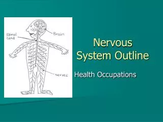
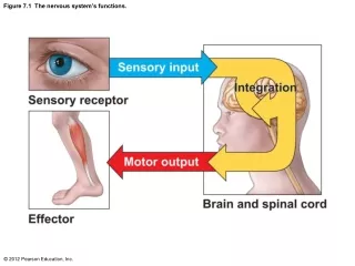
Figure 7.1 The nervous system’s functions. Figure 7.2 Organization of the nervous system. Figure 7.3 Supporting (glial) cells of nervous tissue. Figure 7.3a Supporting (glial) cells of nervous tissue. Figure 7.3b Supporting (glial) cells of nervous tissue.

lwhitehead
Télécharger la présentation

Figure 7.1 The nervous system’s functions.

An Image/Link below is provided (as is) to download presentation Download Policy: Content on the Website is provided to you AS IS for your information and personal use and may not be sold / licensed / shared on other websites without getting consent from its author. Content is provided to you AS IS for your information and personal use only. Download presentation by click this link. While downloading, if for some reason you are not able to download a presentation, the publisher may have deleted the file from their server. During download, if you can't get a presentation, the file might be deleted by the publisher.

E N D

Presentation Transcript


  1. Figure 7.1 The nervous system’s functions.

  2. Figure 7.2 Organization of the nervous system.

  3. Figure 7.3 Supporting (glial) cells of nervous tissue.

  4. Figure 7.3a Supporting (glial) cells of nervous tissue.

  5. Figure 7.3b Supporting (glial) cells of nervous tissue.

  6. Figure 7.3c Supporting (glial) cells of nervous tissue.

  7. Figure 7.3d Supporting (glial) cells of nervous tissue.

  8. Figure 7.3e Supporting (glial) cells of nervous tissue.

  9. Figure 7.4 Structure of a typical motor neuron.

  10. Figure 7.4a Structure of a typical motor neuron.

  11. Figure 7.4b Structure of a typical motor neuron.

  12. Figure 7.5 Relationship of Schwann cells to axons in the peripheral nervous system.

  13. Figure 7.5a Relationship of Schwann cells to axons in the peripheral nervous system.

  14. Figure 7.5b Relationship of Schwann cells to axons in the peripheral nervous system.

  15. Figure 7.5c Relationship of Schwann cells to axons in the peripheral nervous system.

  16. Figure 7.6 Neurons classified by function.

  17. Figure 7.7 Types of sensory receptors.

  18. Figure 7.7a Types of sensory receptors.

  19. Figure 7.7b Types of sensory receptors.

  20. Figure 7.7c Types of sensory receptors.

  21. Figure 7.7d Types of sensory receptors.

  22. Figure 7.7e Types of sensory receptors.

  23. Figure 7.8 Classification of neurons on the basis of structure.

  24. Figure 7.8a Classification of neurons on the basis of structure.

  25. Figure 7.8b Classification of neurons on the basis of structure.

  26. Figure 7.8c Classification of neurons on the basis of structure.

  27. Figure 7.9 The nerve impulse.

  28. Figure 7.10 How neurons communicate at chemical synapses.

  29. Figure 7.11 Simple reflex arcs.

  30. Figure 7.11a Simple reflex arcs.

  31. Figure 7.11b Simple reflex arcs.

  32. Figure 7.11c Simple reflex arcs.

  33. Figure 7.12 Development and regions of the human brain.

  34. Figure 7.12a Development and regions of the human brain.

  35. Figure 7.12b Development and regions of the human brain.

  36. Figure 7.13 Left lateral view of the brain.

  37. Figure 7.13a Left lateral view of the brain.

  38. Figure 7.13b Left lateral view of the brain.

  39. Figure 7.13c Left lateral view of the brain.

  40. Figure 7.14 Sensory and motor areas of the cerebral cortex.

  41. Figure 7.15 Frontal section of the brain showing commissural, association, and projection fibers running through the cerebrum and the lower CNS.

  42. Figure 7.16 Diencephalon and brain stem structures.

  43. Figure 7.16a Diencephalon and brain stem structures.

  44. Figure 7.16b Diencephalon and brain stem structures.

  45. Figure 7.17 Meninges of the brain.

  46. Figure 7.17a Meninges of the brain.

  47. Figure 7.17b Meninges of the brain.

  48. Figure 7.18 Ventricles and location of the cerebrospinal fluid.

  49. Figure 7.18a Ventricles and location of the cerebrospinal fluid.

  50. Figure 7.18b Ventricles and location of the cerebrospinal fluid.

More Related